 PORNPAK KHUNATORN/ISTOCK/GETTY IMAGES PLUS
Treating Patients With Gastroparesis
Oral health professionals should be prepared to help patients with this disorder address the oral signs and symptoms.
This course was published in the March 2021 issue and expires March 2024. The authors have no commercial conflicts of interest to disclose. This 2 credit hour self-study activity is electronically mediated.
EDUCATIONAL OBJECTIVES
After reading this course, the participant should be able to:
- Define gastroparesis (GP) and identify its signs and symptoms.
- Describe the management of GP symptoms.
- Discuss the oral health risk factors associated with GP.
Gastroparesis (GP) is a rare and debilitating gastrointestinal motility disorder that results in the complete or partial paralysis of stomach muscles. In health, the stomach chemically and mechanically digests food, then propels it into the small intestines (gastric emptying) where further digestion and nutrient absorption occur. In the case of GP, impaired muscles impact the stomach’s ability to mechanically digest and propel food into the small intestines, leading to malnourishment and recurrent symptoms of nausea, vomiting, severe upper abdominal pain, early satiety, and bloating.1–4
The diagnosis of GP is determined by the presence of symptoms, lack of stomach obstruction, and delayed gastric emptying; however, the etiology of this disorder is multifaceted.1,2 The stomach muscle impairments associated with GP have been linked to the diagnoses of post-operative gastroparesis and several underlying health conditions including, Parkinson disease, neurological disorders, scleroderma, post-viral infections, and, most frequently, diabetes. While many known factors contribute to the development of GP, more than half of all cases are idiopathic (no known cause).1–7
Due to its subclinical nature, the prevalence of GP is unknown. However, recent data suggest that GP impacts approximately 0.16% of the United States population.8 GP is predominantly observed in women (66% to 80%), and is more prevalent among white people compared to other racial groups.3,8 Though GP can occur at any age, the type of GP is often related to the age of onset:8
- Idiopathic (age < 39)
- Type 1 diabetes (between ages 40 and 59)
- Type 2 diabetes (between ages 60 and 80)
Studies have shown that patients with GP experience a reduced quality of life compared to the general public in addition to high mortality rates. Upward of 10% of those with GP become disabled due to the disorder’s effects.3,9,10
MEDICAL MANAGEMENT/TREATMENT
Currently, there is no known cure for GP. The medical management of the disorder begins by identifying and treating underlying conditions. Other GP treatment strategies include managing symptoms and increasing gastric emptying.4,11 Symptoms and their severity may vary from day to day. This unpredictability makes it imperative for patients and healthcare providers to work closely together while managing the disorder. Treatment strategies include but are not limited to dietary modifications (Table 1), fluid and electrolyte restoration, liquid nutrients (protein shakes and smoothies), administration of antiemetics (anti-vomiting drugs), prokinetics (gastric stimulating drugs), gastric electrical stimulators, and feeding tubes.2,11–13
![TABLE 1. Suggested Dietary Modifications to Alleviate Gastroparesis Symptoms]() ORAL SIDE EFFECTS OF GASTROPARESIS
ORAL SIDE EFFECTS OF GASTROPARESIS

Oral and systemic health are intrinsically related. The oral cavity is often referred to as the mirror to the body because it can reveal signs of disease and nutritional deficiencies, as well as symptoms of poor diet and recreational drug use. The oral cavity is also the start of the gastrointestinal tract and is responsible for initiating the chemical digestion through salivary enzymes and mechanical digestion through mastication. As such, patients with GP need to ensure their tooth structures and salivary glands are intact and function properly to aid their already compromised digestive processes.
As oral disease prevention specialists, dental hygienists are on the frontline when it comes to recognizing the oral manifestations of systemic diseases. Because GP is underdiagnosed, dental hygienists should not only be aware of the disorder’s signs and symptoms, but also its oral manifestations. Dental hygienists are well-positioned to help undiagnosed patients seek appropriate medical care and to collaborate interprofessionally as part of the patient’s healthcare team.
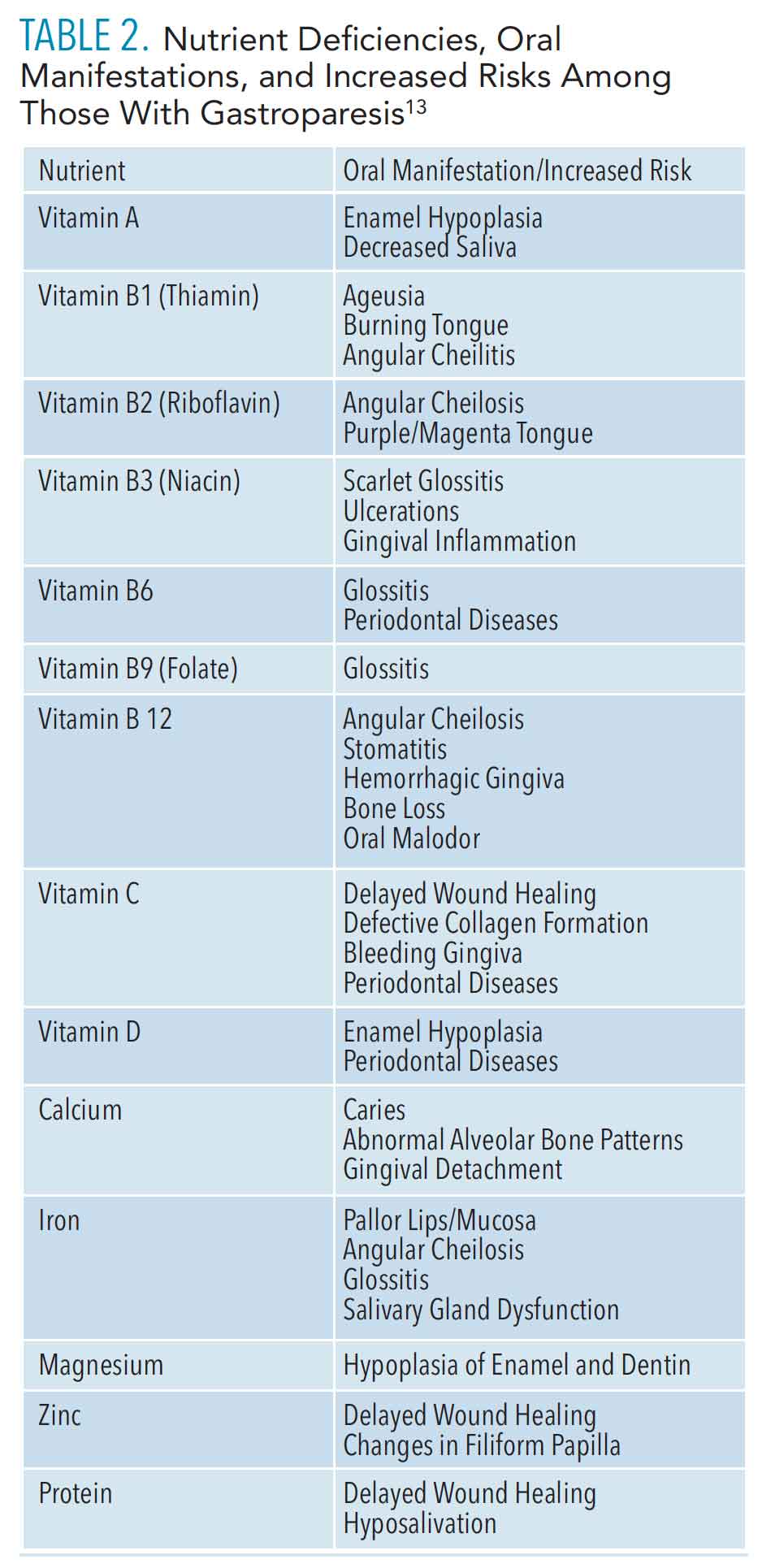
Due to the multifaceted nature of GP, patients are at high risk of oral side effects. Hyposalivation is observed in severe cases of malnutrition or is caused by antiemetic drugs. Decreased saliva flow, along with frequent nausea and vomiting and strict dietary modifications, increase patients’ risk of developing erosive tooth wear (ETW), dentinal hypersensitivity, and dental caries.14–16 Vomiting, dietary restrictions, early satiety, and/or slow gastric emptying rates contribute to malnutrition, which further impacts periodontal health and development of several oral conditions related to vitamin and nutrient deficiencies, including but not limited to, glossitis, angular cheilitis, pallor gingiva, stomatitis, and oral malodor (Table 2).
![table 2]() DENTAL CONSIDERATIONS
DENTAL CONSIDERATIONS
Because patients with GP are susceptible to ETW, dentinal hypersensitivity, and caries, dental hygienists need to understand the similarities and differences of these calcified tooth structure disease processes. All three conditions result from an equilibrium imbalance of tooth mineralization processes and the dissolution of the organic matrix of tooth structures. Demineralization occurs when mineral ions (calcium and phosphate) are removed from hard tissue tooth structures. On the other hand, remineralization is the process of moving minerals, including hydrogen fluoride, back to tooth surfaces. Saliva acts to facilitate the continuous process of demineralization and remineralization. It also protects tooth structures by buffering destructive-acidic pH levels that break down and dissolve tooth surfaces.14,17–19 When patients experience hyposalivation, an imbalance occurs in the demineralization and remineralization processes, increasing the rate of demineralization, often leading to tooth erosion, dentinal hypersensitivity, and/or dental caries.
Dental hygienists must further recognize how the etiologies of ETW, dental hypersensitivity, and dental caries differ. Understanding these differences while comprehensively assessing patients’ oral health and GP statuses, will help clinicians to treat, maintain, and, ultimately, prevent tooth destruction for patients with GP.
ETW is a result of nonbacterial acids (stomach acids) caused by pH fluctuations in the throat and oral cavity as well as other factors, including nutrition and patient behaviors that sensitize teeth to stimuli.18,19 Patients with GP who frequently vomit may present with smooth, saucer-like depressions on the lingual surfaces of their anterior teeth. In severe cases, ETW may extend into the dentin and/or compromise the integrity of dental restorations throughout the mouth. Clinical signs of ETW must be recognized and diagnosed as early as possible so that appropriate, individualized risk management and treatment plans can be swiftly implemented. Interestingly, patients who report vomiting should be instructed to avoid brushing immediately after vomiting and instead swish with water or sodium bicarbonate to help neutralize acids. Additionally, products containing fluoride, casein phosphopeptide-amorphous calcium phosphate (CPP-ACP), and xylitol should be recommended as they effectively buffer salivary pH levels.14 Patients with GP may be unaware of their actual acid intake; best practice dictates using a dietary and behavior diary over 4 days to help adjust eating and drinking habits that can prevent further destruction from ETW.15,20–22
ETW is also a significant risk factor for dentinal hypersensitivity. In health, dentinal tubules are protected by enamel. However, the destructive nature of ETW breaks down enamel, exposing the nerve fibers within dentinal tubules to the oral cavity. Once the tubules are open, stimuli from food, beverages, toothbrushing, and dental care can elicit a painful response that, in severe cases, can negatively impact quality of life.23 To effectively treat patients experiencing dental hypersensitivity, determining what type of stimuli is causing the pain, as well as noting its frequency and severity, is important. Glass ionomer and resin-based composites can be placed in the office to treat localized dental hypersensitivity. Varnishes, toothpastes, and mouthrinses containing sodium fluoride, stannous fluoride, and strontium salts can help prevent generalized symptoms of dentinal hypersensitivity by occluding dentinal tubules, protecting nerve endings from painful stimuli. In-office products containing calcium sodium phosphosilicate, arginine with calcium carbonate, glutaraldehyde hydroxyethyl methacrylate, or potassium binoxalate with nitric acid are also available. Products with oxalates act by decreasing dentinal permeability while occluding dentinal tubules, and potassium nitrate-based products decrease sensitivity by actively depolarizing nerve endings.24 Regardless of treatment modality, oral health professionals need to work closely with the patient to effectively reduce and maintain the pain associated with dental hypersensitivity.
While diminished salivary flow rates are a contributing factor, caries is induced by dental biofilm, resulting in the destruction of mineralized tooth structures.25 Acidogenic bacteria, such as Streptococci mutans, have been implicated in the caries disease process. S. mutans adhere to tooth structures, consume and metabolize fermentable carbohydrates, and produce harmful tooth-demineralizing acids. The frequent ingestion of foods and beverages containing fermentable carbohydrates, along with other risk factors such as poor biofilm control, malnutrition, and systemic conditions, increase the probability of caries development.
Caries risk must be carefully assessed prior to developing a management strategy for patients with GP using the “caries management by risk assessment” (CAMBRA) criteria. CAMBRA is a process that rates caries risk as low, medium, or high. It involves carefully collecting information from the patient’s medical and dental history, conducting a complete clinical examination, detecting and identifying carious lesions that may be reversed or prevented, and developing a customized treatment plan for the patient that includes any professional treatments or over-the-counter measures to help prevent the development of future dental caries.26 Products containing fluoride, CPP-ACP, and/or xylitol inhibit acidogenic bacterial growth, buffer salivary pH, and stimulate salivary flow rates. When present in saliva, fluoride and CPP-ACP aid in the remineralization process. Fluoride and ACP both act by creating a crystal matrix on tooth surfaces that attract calcium and phosphate ions. CPP saturates saliva and plaque biofilm with calcium and phosphate, increasing tooth remineralization.14 As patients with GP often experience malnutrition, they are more likely to be deficient in calcium and phosphorous, making casein-based products important for the prevention and maintenance of dental caries.
ROLE OF THE DENTAL HYGIENIST
While little is known about providing dental care specifically for patients with GP, dental hygienists can effectively address the disorder’s common oral side effects. Patient care should center on interprofessional collaboration, oral health risk assessments, and individualized treatment and management plans. Therefore, dental hygienists must be prepared to collaborate with members of the patient’s medical team, as well as dietitians. Understanding the full extent of the patient’s diagnosis and/or underlying health condition, current GP symptoms and status, dietary modifications, and medications will help the dental hygienist to comprehensively develop a care plan.
Professional interventions should first focus on treating any underlying health conditions. For example, if a patient presents with type 2 diabetes-associated GP, dental hygiene diabetes standards of care would be followed first and carefully documented in the patient’s chart. Any diabetes-associated nutritional counseling should uphold the treatment plan of the patient’s nutritionist and/or medical team.
Dental hygienists should also offer preventive care to not only help improve the patient’s overall quality of life, but to manage risk factors for oral disease. To achieve this, dental hygienists should update risk assessment and management plans at each recare appointment. Appointment intervals should be based on current oral health and GP statuses.
For patients experiencing hyposalivation, salivary gland function should be assessed routinely at dental visits.15,27 Treatment of hyposalivation should include increased frequency of water consumption; saliva substitutes, such as gels, sprays, or mouthrinses; and xylitol chewing gum. Research shows that saliva substitutes can increase remineralization due to the increased viscosity of the product, increasing the mechanical protection of the tooth surface.28
Patients at high risk for ETW, dental hypersensitivity, caries, and hyposalivation should use extra-soft toothbrushes, fluoride- and/or casein-based products, saliva substitutes, and products containing xylitol. Patients should avoid brushing their teeth immediately after vomiting and/or eating and drinking, and instead swish with water or hydrogen peroxide to neutralize oral pH levels. When treating patients with ETW, dental hygienists should avoid using abrasive prophy pastes and powders to reduce the risk of further tooth destruction. Concerns surrounding nutrient-related oral manifestations should be addressed collectively with patients and their medical team.
CONCLUSION
In conclusion, GP is a rare and often debilitating disorder with no known cure. Due to the complex nature of GP, patients are at high risk of experiencing several adverse oral conditions including but not limited to hyposalivation, ETW, dentinal hypersensitivity, dental caries, and oral manifestations specifically related to malnutrition. Though evidence-based dental protocols are developing on this topic, dental hygienists are well prepared to critically assess and comprehensively manage the common oral side effects of GP. Oral healthcare provided should be performed collectively with the patient, as well as the dental and medical healthcare teams in order to effectively improve the patient’s oral health status and overall quality of life.
REFERENCES
- American College of Gastroenterology. Gastroparesis. Available at: gi.org/topics/gastroparesis. Accessed February 16, 2021.
- Camilleri MD, Parkman HP, Mehnaz SA, et al. Clinical guideline: management of gastroparesis. Am J Gastroenterol. 2013;108:18–38.
- Lacy BE, Crowell MD, Mathis C, et al. Gastroparesis: quality of life and health care utilization. J Clin Gastroenterol. 2018;52:20–24.
- Mayo Clinic. Gastroparesis Symptoms and Causes. Available at: mayoclinic.org/diseases-conditions/gastroparesis/symptoms-causes/syc-20355787. Accessed February 16, 2021.
- Rey E, Choung RS, Schleck CD, et al. Prevalence of hidden gastroparesis in the community: the gastroparesis “iceberg.” J Neurogastroenterol Motil. 2012;18:34–42.
- Hasler ML. Gastroparesis: symptoms, evaluation, and treatment. Gastroenterol Clin N. 2007;36:619–647.
- Grover M, Farrugia G, Stanghellini V. Gastroparesis: a turning point in understanding and treatment. Gut. 2019;68:2238–2250.
- Syed AR, Wolfe MM, Calles-Escandon J. Epidemiology and diagnosis of gastroparesis in the United States. J Clin Gastroenterol. 2020;54:50–54.
- Hyett B, Martinez FJ, Gill BM, et al. Delayed radionucleotide gastric emptying studies predict morbidity in diabetics with symptoms of gastroparesis. Gastroenterol. 2009;137:445–452.
- National Organization for Rare Disorders. Gastroparesis. Available at: rarediseases.org/rare-diseases/gastroparesis. Accessed February 16, 2021.
- Guideline Central. Clinical Guideline: Management of Gastroparesis Clinical Practice Guidelines. Available at: guidelinecentral.com/summaries/clinical-guideline-management-of-gastroparesis/#section-society. Accessed February 16, 2021.
- Parkman HP, Hasler WL, Fisher RS. American Gastroenterological Association medical position statement: diagnosis and treatment of gastroparesis. Gastroenterol. 2004;127:1589–1591.
- United States Department of Agriculture. Methods and Application of Food Composition Laboratory. Available at: ars.usda.gov/nutrientdata. Accessed February 16, 2021.
- Farooq I, Moheet IA, Imran Z, Farooq U. A review of novel dental caries preventive material: casein phosphopeptide-amorphous calcium phosphate (CPP-ACP) complex. King Saud University Journal of Dental Sciences 2013;4:47–51.
- Yoskikawa H, Furuta K, Ueno M, et al. Oral symptoms including dental erosion in gastroesophageal reflux disease are associated with decreased salivary flow volume and swallowing function. J Gastroenterol. 2012;47:412–420.
- Buzalaf M, Magalhaes A, Rios D. Prevention of erosive tooth wear: targeting nutritional and patient-related risks factors. Br Dent J. 2018;224:371–378.
- Furgeson D, Pitts E. Saliva’s role in remineralization. Dimensions of Dental Hygiene. 2018;16(5):26–29.
- Frese C, Frese F, Kuhlman S, et al. Effect of endurance training on dental erosion, caries, and saliva. Scand J Med Sci Sports. 2015;25:319–326.
- Mulic A, Bjorg Tveit A, Songe D, Silvertsen H, Skaare A. Dental erosive wear and salivary flow rate in physically active young adults. BMC Oral Health. 2012;12:8.
- Lussi A, Carvalho T. Erosive tooth wear: a multifactorial condition of growing concern and increasing knowledge. Monogr Oral Sci. 2014;25:1–15.
- Lussi A, Hellwig E. Risk assessment and causal preventive measures. Monogr Oral Sci. 2014;25:220–229.
- Alaraudanjoki V, Laitala M, Tjaderhane, L., et al. Influence of intrinsic factors on erosive tooth wear in a large-scale epidemiological study. Caries Res. 2016;50:508–516.
- O’Toole S, Bartlett D. The relationship between dentine hypersensitivity, dietary acid intake and erosive toothwear. J Dent. 2017;67:84–87.
- Miglani S, Aggarwal V, Ahuja B. Dentin hypersensitivity: recent trends in management. J Conserv Dent. 2010;13:218–224.
- Bowen W, Burne R, Wu H, Koo H. Oral biofilms: pathogens, matric and polymicrobial interactions in microenvironments. Trends Microbiol. 2018;26:229–242.
- Fetherstone J, Chaffee B. The evidence for caries management by risk assessment (CAMBRA). Adv Dent Res. 2018;29:9–14.
- Navazesh M, Kumar S. Measuring salivary flow: challenges and opportunities. J Am Dent Assoc. 200;139:35S–40S.
- Aykut-Yetkiner A, Wiegand A, Attin T. 2014. The effect of saliva substitutes on enamel erosion in vitro. J Dent. 2014;42:720–725.
From Dimensions of Dental Hygiene. March 2021;19(3):32-35.
