Digital Technology Integration
The following strategies can help oral health professionals implement new technologies into daily clinical practice.
This course was published in the September 2017 issue and expires September 2020. The authors have no commercial conflicts of interest to disclose. This 2 credit hour self-study activity is electronically mediated.
EDUCATIONAL OBJECTIVES
After reading this course, the participant should be able to:
- Describe the digital solutions currently available in the oral health care setting.
- Discuss the accuracy of digital impressions.
- Identify the return on investment for digital solutions.
- Explain the difference between open and closed files.
Digital technology plays an influential role in the delivery of oral health care, and treatment is evolving rapidly as a result of ongoing advances. Digital radiology and photography, for example, help improve diagnostic capabilities and outcomes. In addition, many routine dental processes have been positively impacted by digital technology, including communication and information handling, claims processing and insurance reimbursement, and practice management through metrics tracking. Initially, digital dentistry focused on restoration of teeth with inlays, onlays, crowns, and veneers. As the technology has developed, additional applications have been introduced, including fixed partial dentures (FPDs), removable appliances, and orthodontic treatment planning and appliance fabrication. These systems also have application for diagnostic implant planning and surgical guide fabrication, as well as abutment design and fabrication. This expanded capability across a range of services has many oral health professionals wondering how to integrate this technology into daily practice.
Terms such as digital technology, digital dentistry, digital imaging, and computerized dentistry tend to be used interchangeably, with a common meaning of digital imaging and computer-aided design/computer-aided manufacturing (CAD/CAM). These modalities may be thought of as a clinical workflow involving three specific sequences or functions, and a number of articles have described the process in detail.1–4 The first function is to record the intraoral condition to computer-based software. This requires a camera, wand, or intraoral scanner, and is often referred to as digital imaging or taking a digital impression. Once the data file has been created on the software, the second sequence involves using a program to plan or design a device—such as a restoration, abutment, implant surgical guide, or bite splint. Once it has been designed, the third sequence is to manufacture the device. Subtractive milling or three-dimensional (3D) printing are the primary techniques used in dental CAM processes. Subtractive milling involves cutting or grinding away excess material from a premanufactured block of material, such as porcelain or zirconia, leaving the desired volumetric shape. Conversely, 3D printing involves additive layers of a resin-based material to create the desired volumetric shape.
With the increasing array of clinical applications, the challenge for oral health professionals is deciding how to optimally integrate CAD/CAM technology into practice. Focusing on the equipment initially can be confusing, especially when determining which devices can perform which functions—and at what cost. When considering purchase decisions, one obvious element of success with CAD/CAM technology is that it is only useful if it is routinely applied in treatment. To successfully integrate CAD/CAM technology into practice, clinicians must start with an understanding of what functions are of value now, as well as the capabilities they seek for future expansion of services.
DIGITAL SOLUTIONS
FIGURE 1A. Digital impressions provide crisp detail.
One approach to making sense of digital dentistry is to divide the equipment into two categories, based on which function in the CAD/CAM workflow is desirable. The first category is digital imaging or digital impression systems. This modality focuses on the first sequence of the clinical workflow: accurately recording the intraoral condition to a computer, and transmitting the digital files to a laboratory for design and fabrication of the desired device. Software included with digital impression systems, however, does not allow operators to design or plan any sort of device or restoration. It is primarily administrative software that allows for patient identification and case transmission to a dental laboratory for design and fabrication. At best, clinicians can see immediate feedback on the tooth preparation.
The second broad category is chairside CAD/CAM systems, which combine all three functions in the dental office. These systems also record intraoral scans, but include a program for designing restorations and a milling unit to fabricate the restoration. They are designed to leverage the efficiency of a single-appointment procedure to the benefit of both the patient and practice.
Digital impression systems have evolved from single image capture technology using a reflective powder to full color video recording. Not only is video recording helpful during the design and fabrication processes, it is also an effective way to educate patients on the condition needing treatment.
Initial CAD/CAM units included a computer, monitor, and camera in a stand-alone cart design that could be unwieldy to move around the office. Today, manufacturers offer modular options that allow the cameras to be connected to computers, with the monitor more advantageously positioned in the operatory. Although reported to be a more streamlined setup, the laptop still must be located adjacent to the operating field for direct access to the imaging device on either an adjacent counter or cart.
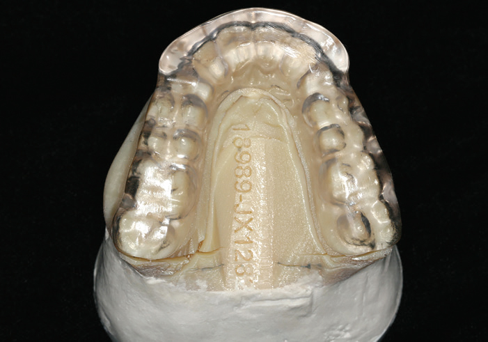
FIGURE 1B. Maxillary bite splint processed on the digital model.
For many clinicians, the learning curve is a significant concern regarding the implementation of CAD/CAM technology into practice. Compared to chairside CAD/CAM systems, the curve is generally shorter and easier for digital impression technology. Digital imaging system software is fairly easy to learn, as it focuses primarily on case identification and information input to electronically transmit the case to the lab with prescription instructions. While chairside CAD/CAM technology can also function as a digital impression system, it presents a steeper curve due to the need to become familiar with the design techniques, fabrication functions, and tools available in the software.
DIGITAL IMPRESSION ACCURACY
Compared to digital scans, one limitation of conventional impressions is the degradation of accuracy when multiple pours are made from the same impression. Digital impressions avoid many of the potential problems of conventional impressions, with the added advantage that models can be fabricated with equal accuracy an unlimited number of times, as the digital file does not undergo any loss of accuracy as it is repeatedly processed.
Some clinicians have expressed concerns about the accuracy of the restoration outcome when using digital or conventional impression techniques. An in vitro study investigated the marginal and internal fit of FPDs fabricated using conventional or digital impressions.5 A CAD program was used to digitally measure margin gap, internal adaptation, and cervical discrepancies between the FPDs. The results showed no significant differences in the marginal gap between restorations made using conventional or digital impressions; however, the internal and cervical discrepancies were less for restorations fabricated from digital impressions.
A more extensive in vitro study compared the accuracy of ceramic crowns designed and fabricated based on various digital impression systems and traditional impression techniques.6 Conventional impressions were made of a single crown preparation on a master model using two-step and single-step putty wash impression techniques. Digital impressions were made of the master model crown preparation using three systems. The mean margin fit of crowns was reported as 48 ± 25 microns for system one, 30 ± 17 microns for system two, and 41 ± 16 microns for system three. By comparison, it was reported as 33 ± 19 microns for single-step putty wash technique and 60 ± 30 microns for the two-step putty wash technique. There was no significant difference in the margin fit or internal adaptation of the crowns using any of the techniques.
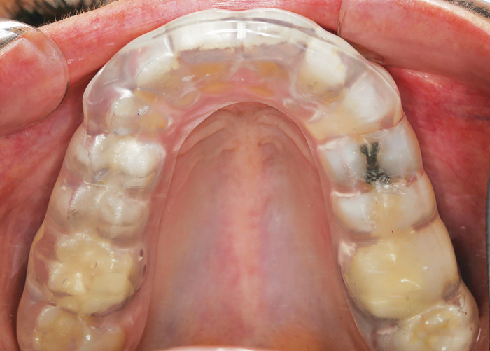
FIGURE 1C. Maxillary bite splint fabricated from a fullarch scan.
In a randomized clinical study, researchers compared the margin fit of two types of zirconia crowns fabricated from either conventional or digital impressions.7 A vinyl polysiloxane impression and digital impression were taken for each of 50 crown preparations. Crowns were made for each preparation using the two impression techniques. Each crown was measured intraorally for margin fit and internal adaptation using a replica technique. The crowns made with the digital impression demonstrated a margin fit (51.45 ± 18.59 microns) that was superior to those made with a conventional impression technique (78.62 ± 25.62 microns).
From a learning curve perspective, clinicians who are considering digital capabilities may wish to incrementally integrate CAD/CAM technology into practice. For example, a digital impression system could be procured initially, and, as comfort with digital scanning increases, other chairside functions may be added. To date, this has only been possible with chairside CAD/CAM systems. The acquisition unit may be purchased without the milling unit, as these systems also function as digital impression systems. The milling unit could be added at a later date.
A trend in digital imaging systems is the ability to function with third-party software and milling units, in essence moving to chairside CAD/CAM systems. A chief difference between chairside CAD/CAM configurations using equipment from several manufacturers and a system from a single manufacturer is the variety of clinical outcomes afforded. The single-manufacturer chairside CAD/CAM systems can generally process a wider variety of materials, including glass ceramics, nanoceramics, resin-based materials, lithium disilicate, and full contour zirconia. The extended digital impression/CAD/CAM systems may be limited to one or two specific types of materials and restorations.
RETURN ON INVESTMENT
Chairside CAD/CAM systems are designed to leverage the efficiency of the digital workflow in a single appointment for indirect restorations that usually require multiple appointments. This eliminates the need for a provisional restoration, as well as a second appointment to deliver the final restoration. This benefits both the patient (in terms of convenience) and the practice (in terms of productivity). That said, purchasing a chairside CAD/CAM system represents a sizable capital investment, so careful consideration of the return on investment is critical to the decision-making process.
When planning such a purchase, oral health professionals will generally consider what CAD/CAM technology will replace in the practice. While digital technology is not a replacement for impression material, it should be evaluated for how it may transform the practice. For example, the digital impression process is often compared with the time and cost of making a vinyl polysiloxane impression. Although conventional and digital impressions can be used to provide accurate restorations, the “replacement approach” overlooks one of the most significant advantages chairside CAD/CAM technology brings to a practice. The question becomes, “What is it worth to prepare and deliver a crown in a single appointment?” This is the transformation CAD/CAM technology offers—the ability to deliver a significantly improved patient experience in a more efficient workflow for the patient and practice.
OPEN VS CLOSED FILES
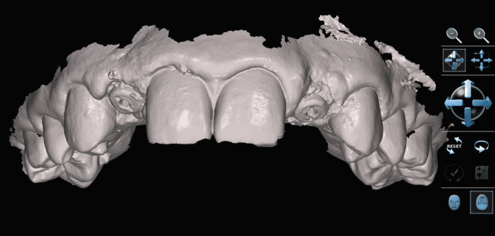
FIGURE 2A. Digital technologies are invaluable for implant planning and delivery, as seen with these encode abutments for implants #7 and #10.
Files from various types of digital systems have generally been labeled as “open” or “closed” files, which refers to what software and equipment may use the data. Open implies that the digital file can be exported from the intraoral scanning device and be read by any CAD software. Closed files generally refer to digital data that are managed by a single manufacturer, such as a chairside CAD/CAM system in which the camera, software, and milling unit are from the same manufacturer. In other words, closed means the digital file is proprietary and must be unlocked prior to the file being opened by CAD software from another manufacturer. At this time, there is no universally accepted file format that manufacturers conform to for digital systems. Dental laboratories may prefer open files to avoid the need for multiple software versions or multiple versions of equipment to handle the various types of files that practices may send for processing.
A system’s digital file architecture is relevant due to the financial implications of unlocking a file. Some manufacturers claim to have an open architecture, but require the file to be sent through a portal to enjoy the full capabilities of an open file. Although the file will be open architecture, there is often a fee associated with being unlocked. Additionally, some systems require the purchase of software licenses to unlock the capabilities in their CAD software. The open and closed file labels have evolved to become relatively inconsequential. Rather than evaluating systems based on how the digital files are labeled, the focus should be on the clinical applications and capabilities the office seeks.

FIGURE 2B. A stereolithography-processed model from the scan, with fabricated custom abutments and crowns.
As noted, the initial applications for chairside CAD/CAM systems were to produce single-tooth restorations, but now some offer the ability to fabricate short-span FPDs, adhesively bonded FPDs, and provisional restorations in the dental office. One of the more advantageous advances is the ease and efficiency with which full-arch scans can be recorded. This has led to improvements in managing more complex occlusal schemes and movements through virtual articulators that can simulate lateral and protrusive guidance. Full-arch scans can also be used in the fabrication of tooth-borne appliances, such as bite splints (Figure 1A to Figure 1C). It has also led to more orthodontic applications, especially in the area of clear aligner therapy.
INTEGRATING CONE-BEAM IMAGING
Another expanding use of in-office digital technology involves dental implants. Cone-beam computed tomography (CBCT) has become a preferred preoperative 3D diagnostic tool for implant treatment planning. In addition, CAD/CAM surgical guides can be created to facilitate precise implant placement. A number of chairside CAD/CAM systems can integrate the design file from their software program with the file from the CBCT to support a sophisticated, prosthetic-influenced treatment approach. Use of these modalities precludes the need to do a traditional diagnostic wax-up or diagnostic setup, as it can be accomplished virtually through the CAD software and digital file integrated with the CBCT file. This allows for surgical guides to be ordered through the digital planning system or designed and milled in-office.
The second implant-specific application is design and fabrication of provisional and definitive custom abutments and restorations (Figure 2A to Figure 2C). The use of scan posts and scan bodies in lieu of traditional fixture-level impression copings records the implant position and alignment within the dentition. This can be matched with overlying soft tissue scans for development of the custom abutment contours, as well as crowns.8
CONCLUSION
FIGURE 3C. Delivery of custom abutments and crowns for implants #7 and #10, with conservative composite restorations on #8 and #9.
As more configurations and capabilities are introduced, clinicians can easily be overwhelmed when considering the optimal combination to integrate into practice. Digital impression systems leverage the efficiency, convenience, and accuracy of the digital restorative workflow, but without the need to integrate in-office design and fabrication processes. A chairside CAD/CAM system is capable of fabricating restorations, FPDs, provisional restorations, implant abutments, and surgical guides, as well as function as a digital impression system for use in communicating with laboratories. Consequently, the choice of a system should be based on the clinical applications appropriate for the practice, as well as services the office wishes to add in the future.
Digital impression and chairside CAD/CAM systems have been validated in multiple independent studies. Additionally, more than 30 years of in vivo data confirm that chairside CAD/CAM all-ceramic restorations are an esthetic, durable, and predictable option that can preserve tooth structure while leveraging the benefits of adhesive dentistry. Ongoing advances in digital technology and all-ceramic materials are encouraging oral health professionals to take the leap from considering whether CAD/CAM technology fits their practice to deciding how it can best be integrated into existing workflows.
REFERENCES
- Beuer F, Schweiger J, Edelhoff D. Digital dentistry: An overview of recent developments for CAD/CAM generated restorations. Br Dent J. 2008;204:505–511.
- Birnbaum NS, Aronson HB. Dental impressions using 3D digital scanners: Virtual becomes reality. Compend Contin Educ Dent. 2008;2:494–505.
- Fasbinder DJ. Digital dentistry: Innovation for restorative treatment. Compend Contin Educ Dent. 2010;31:2–11.
- Fasbinder DJ. Using digital technology to enhance restorative dentistry. Compend Contin Educ Dent. 2012;33:666–677.
- Svanborg P, Skjerven H, Carlsson P, Eliasson A, Karlsson S, Ortorp A. Marginal and internal fit of cobalt-chromium fixed dental prostheses generated from digital and conventional impressions. Int J Dent. 2014;2014:534382.
- Seelbach P, Brueckel C, Wöstmann. Accuracy of digital and conventional impression techniques and workflow. Clin Oral Invest. 2013;17:1759–1764.
- Fasbinder DJ, Neiva GF, Dennison JB, Heys D, Heys R. Evaluation of Zirconia Crowns Made From Conventional and Digital Impressions. Available at: researchgate.net/publication/266778920_Evaluation_of_Zirconia_Crowns_made_from_Conventional_and_Digital_Impressions. Accessed August 9, 2017.
- Wittneben JG, Wright RF, Weber HP, Gallucci GO. A systematic review of clinical performance of CAD/CAM single-tooth restorations. Int J Prosthodont. 2009;22:466–471.
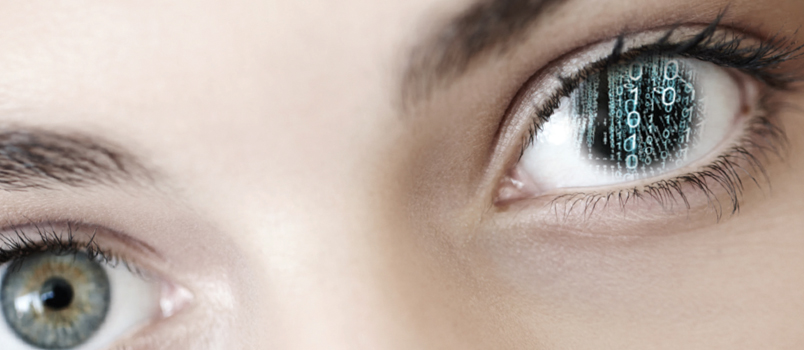
Featured photo by GLOBALSTOCK/E+/GETTY IMAGES PLUS
From Dimensions of Dental Hygiene. September 2017;15(9):36-39.
