The New Frontier
The incorporation of telehealth technologies into forensic odontology significantly improves the victim identification process.
This course was published in the January 2014 issue and expires January 2017. The authors have no commercial conflicts of interest to disclose. This 2 credit hour self-study activity is electronically mediated.
EDUCATIONAL OBJECTIVES
After reading this course, the participant should be able to:
- Discuss the application of technology in the process of victim identification in multiple fatality incidents.
- Detail the role of dental professionals in forensic odontology.
- Review the importance of the human dentition in the victim identification process.
- Explain how telehealth technology has improved the field of forensic odontology.
Forensic odontology—the branch of forensic medicine that positively identifies deceased individuals through methods of meticulous data collection, physical examination, and documentation—is a viable component of telehealth. As the application of digital data and telecommunication strategies, telehealth enables the provision of remote health care, health education for patients and professionals, public health, and health administration. During mass fatality incidents, forensic teams use telehealth technologies to collect, organize, interpret, and document data that assist in the victim identification process.
The term forensic odontology was first defined in 1970 by Keiser-Neilson1 as the “branch of forensic medicine which, in the interest of justice, deals with the proper handling and examination of dental evidence and with the proper evaluation and presentation of the dental findings.” This scientific process has been applied in investigations of murder, accidental death, assault, abuse, and multiple fatality incidents.2,3
The use of forensic odontology dates back to 49 AD.2 Modern technology, however, has thrust forensic odontology into the realm of telehealth. Traditional methods of positive identification are being supplanted by improved technologies, and once complex challenges have been effectively addressed with convenient strategies that ensure accuracy and expedite the victim identification process.4
Rapid availability of data is crucial during large-scale multiple fatality incidents, varying degrees of which have been experienced across the globe, often involving victims from several different nations.5,6 The 2004 Indian Ocean earthquake and tsunami, for example, claimed 250,000 lives from 35 countries.6 During this multiple fatality incident, obtaining ante-mortem (before death) dental records from international countries was challenging. Once attained, data—riddled with varying charting symbols and international languages—proved difficult to understand.7 Telehealth is mitigating some of these problems, specifically in the use of electronic dental records, wireless communication, computer hardware and software, digital photography and radiography, virtual autopsy, and three-dimensional computed tomography (CT). Dental hygienists should be familiar with these concepts, as they are responsible for the documentation of dental records.8
ANTE-MORTEM COLLECTION
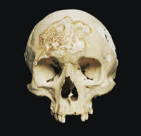
Dentition is resilient to decomposition and extreme heat, and the unique bony structures surrounding it are often the only clues needed to positively identify a victim.9–11 Identity conclusions are made by comparing collective ante-mortem and post-mortem dental data. Forensic odontologists help satisfy legal, societal, and family needs by certifying the identities of victims, and by helping return victims’ remains to their family for proper final arrangements.12
Family members, when assisting in identifying a loved one, may be asked to provide the names and contact information for the victim’s dentist and health care providers, as well as to share videos and photographs that demonstrate the victim’s dental and facial characteristics. The dental office will share a series of ante-mortem data that include written documentation; charts; diagrams; radiographs; clinical photographs and videos; study models; dental materials used; referral letters; orthodontic treatments; test results; laboratory prescriptions; and medical, dental, and social histories.13 Electronic health records and other digitized portions of these items can be wirelessly submitted to authorities, which helps speed the process of identification.6 Collected data are downloaded and analyzed in an identification database.5
Relevant privacy regulations for obtaining a victim’s dental records apply regardless of whether they are of paper or electronic origin. Under the Health Insurance Portability and Accountability Act, the dentist is expected to comply with law enforcement authorities who properly present a warrant, court order, subpoena, or administrative request for a patient’s dental records.14
POST-MORTEM COLLECTION
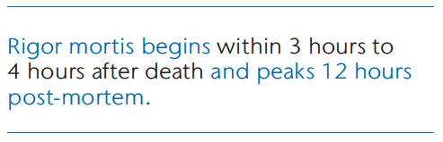
Post-mortem evidence collection begins in the location where the bodily remains were discovered, and it continues in a temporary morgue. Prior to decomposition, fingerprints, facial photographs, intraoral photographs, dental radiographs, and documentation of descriptors (birthmarks, tattoos) are collected. Time is of critical importance; once rigor mortis begins, physical manipulation of the oral cavity proves difficult. Rigor mortis begins within 3 hours to 4 hours after death and peaks 12 hours post-mortem.15 After this point, the mandible may be removed to conduct an intraoral examination or take dental radiographs.5 Removing any part of a deceased body, however, is taboo in some religions.16,17 For this reason, forensic odontology experts work quickly where the body is found to ensure necessary data are collected.
Timely post-mortem analysis employs advanced technologies that allow for accurate data acquisition and collection via portable and digital devices.4 Such advances include digital photography, digital radiographs from portable X-ray units, virtual autopsy, and facial reconstruction (Figure 1) capabilities through three-dimensional CT software. These tools can be used where the body is discovered, in a facility serving as a centralized forensic team information technology center, in a temporary morgue, or as part of a mobile forensic team vehicle.
DIGITAL PHOTOGRAPHY
Photography used in forensic odontology can be performed with a high-quality digital single-lens reflect (SLR) camera, which allows for the lens to be changed; offers multiple exposure settings; and delivers high-resolution imagery—all of which aid in capturing quality evidence.18 Cameras equipped with global positioning system (GPS) tracking tag the coordinates of the location where the victim was found as photos are taken.19 GPS coordinate tags are useful as they enable the forensic team, which may need to gather further evidence, to visit the exact location of the incident at a later date.
Extraoral and intraoral photos should be taken for each victim. If the body of an unknown individual is in good condition, a family member may be able to assert positive identification from a quality full-face photograph. Such assertions, while not scientific or valid for legal purposes, can direct the forensic team in the right direction and often initiate an ante-mortem and post-mortem data comparison.5
Forensic odontologists, to aid in the victim identification process, utilize close-up photos of the victim’s lip prints (cheiloscopy).20 Additionally, post-mortem intraoral photos taken of the palatal rugae pattern can be compared to ante-mortem intraoral photos/dental models using palatal rugae comparison software.21 Lip prints and rugae patterns, like fingerprints, are unique to each individual.9,20–23
DIGITAL RADIOGRAPHY
Digital radiography with portable units is the preferred method for forensic odontology. The digital capture of dental radiographs and other forms of imaging ensures that the necessary protocols regarding chain of evidence and data storage are followed.19 Digital images are magnified for image-spot scrutiny. This requires clear acquisition of the cementoenamel junction outline, pulpal outline, detailed root apex contour, and restoration margin discriminations—all of which are essential to ante-mortem and post-mortem comparisons.9,24
The use of digital radiography also has important logistical benefits. First, it eliminates the need for the forensic team to keep developer and fixer solutions on hand, increasing convenience. Another benefit of digital radiography is that it limits the need for moving fragile bodily remains to another site in order to expose dental radiographs. Disturbances during the transportation process can result in physical deterioration of the remains, therefore compromising the ability to obtain further evidence.25 Portable X-ray units, which are small and lightweight, can be easily transported and used on-site—eliminating the need for access to facilities with fixed-wall units.24 The convenience of a portable unit makes it more likely that radiographs of the victim will be captured before rigor mortis sets in.
VIRTUAL AUTOPSY
Incision-free autopsies may be conducted when combining CT scanning and magnetic resonance imaging (MRI). This autopsy technique is referred to as a “virtual autopsy” and produces a three-dimensional reconstruction of the body, including oral structures, for detailed forensic examination.17 A multidisciplinary team in Switzerland capitalized on this technology by developing the Virtopsy® project—a robotic system guided by three-dimensional surface scanning and computer software programs that allow for precision autopsies of cadavers to be performed with minimal physical damage to the body.26 Virtual autopsy allows fragile, conditioned bodies to remain intact while obtaining detailed data not detectable during traditional autopsy.17
The process begins by inputting collected imagery data into a software program. Photogrammetry and triangulation aided by a digital SLR camera and optical surface scanner wirelessly transmit images to the software. The software calculates millions of surface points on the skin, and a robotic arm—instructed by a navigation system—performs the requested task.16,17 The surface point calculations make it possible for the robot to detect appropriate distance from the skin surface during the examination. The software coordinates the procedure, automates the autopsy workflow, and compiles imagery data to produce the resulting three-dimensional virtual model of the body for examination. Planning and navigation of the robot can be commanded by experts who are off-site, which is helpful during mass fatality incidents that occur in remote locations.26
Virtual autopsy technology is costly, which limits its widespread use. The National Research Council discussed the benefits of virtual autopsy in a 2009 paper, “Strengthening Forensic Science in the United States: A Path Forward,”4 which proposed reform in the forensic sciences field. Forensic dental team members should be aware of the benefits of virtual autopsy due to the head and neck images that it can produce. Use of virtual autopsy eliminates the challenges posed in taking intraoral photos and radiographs when rigor mortis has set in, enhancing the ability to gain sufficient dental evidence.13
THREE-DIMENSIONAL CRANIOFACIAL RECONSTRUCTION
Three-dimensional craniofacial reconstruction recreates a recognizable ante-mortem likeness. This artistic rendering is particularly important for identification when there is no known source of ante-mortem information for skeletonized victims.27 Craniofacial reconstruction can be rendered from traditional methods of an artist’s clay model or three-dimensional CT. According to Pittayapat et al,5 clay models are subjective in nature, whereas CT reconstructions are considered “consistent and objective.” Computerized models are also rendered quickly for transfer to officials who may use the image to publicize the case in hopes that a family member will come forward to identify the victim.
Regardless of the craniofacial reconstruction method used, forensic odontology’s examination of the skull can offer evidence of age estimation to help improve the accuracy of the model’s appearance. Examining the teeth for exfoliation patterns is helpful in estimating the age of young victims who still have all primary teeth or mixed dentition. Victims without primary teeth are assumed to be older than a minimum age. Also, characteristics of secondary teeth can give clues about the victim’s stage of adulthood by patterns seen in the “enamel wear, incremental cementum lines, root translucency, secondary dentin formation, volumetric pulp tooth ratios, and periodontal attachment.”5
DENTAL IDENTIFICATION HARDWARE AND SOFTWARE
Mass fatality incidents can occur anywhere. In the aftermath of a disaster, the forensic team will need a temporary technology center for collection and organization of ante-mortem and post-mortem data.3 The hardware and software components for the technology center vary and depend on the characteristics of the disaster and the needs or preferences of the forensic team.
The Disaster Mortuary Operational Response Team (DMORT) is one of the five teams that comprise the federal National Disaster Medical System (NDMS), which supports the United States’ medical response capability. DMORT offers mortuary assistance in the case of a mass fatality incident or cemetery-related incident. DMORT, in addition to the Unified Victim Identification System (UVIS)—a New York-based online database designed to assist in the handling of mass fatalities—recommend that all forensic teams have access to computers or laptops equipped with a CD drive/burner, USB drive, Windows XP, broadband Internet connectivity, a USB cable to connect digital cameras, scanners, laser color printers, telephone/fax capabilities, and Web cameras technology to effectively transfer and archive data.28
Dental identification software used by forensic odontology teams enables collected data to be electronically catalogued and filtered so that ante-mortem and post-mortem information can be easily compared. The American Board of Forensic Odontology (ABFO) and DMORT both utilize the WinID software system.3 WinID is a paperless dental identification system that contains digitized ante-mortem and post-mortem dental charting, radiographs, and photographs, and enables specific comparisons to be made on a tooth-by-tooth basis.3 Similar international software options include DVI System International® (used by Interpol, the world’s largest international police organization) and DAVID®, which is popular in Australia.5
In addition to cataloguing ante-mortem and post-mortem dental data, the UVIS database contains information about reported missing persons.28 Utilizing the catalog to search for matches is easy and efficient because the data are centralized, organized, and readily available for retrieval. Additionally, UVIS features let users select their preferred language, allowing global users not only to access but understand the data on file. This is especially helpful during disasters with mass fatality incidents that include victims from multiple nations.19,29
DENTAL CODING SYMBOLS DATABASE
Due to previous difficulties in collecting ante-mortem dental charting, forensic odontologists are advocating for a universal dental coding symbols database.9,29 Currently, dental charting from ante-mortem oral health professionals is subject to the dentist’s preferred annotation style. Additionally, symbol coding and dental terminology vary by country, creating the difficult and time-consuming task of decoding and interpreting ante-mortem information. Such challenges force forensic odontologists to decode the information prior to entering it into the identification software, so that comparative analyses can be made. One of the primary challenges in decoding the information is that confusing notations or illegible handwriting may be misinterpreted, which can result in identification errors.7
The Forensic Dental Symbols© and Dental Encoder© Database was created to resolve decoding problems. This system is compatible with Interpol’s disaster victim identification form and allows for international use of uniform graphic symbols and nomenclature.7 If the system were adopted by all clinicians, it would eliminate the need to interpret ante-mortem dental charting because each chart received would be in the correct format.29 Uniformity of the information would eliminate interpretation errors and save valuable time. At present, it is not a requirement for dental professionals to abide by the rules of uniform dental coding in patient records, but the ABFO is calling for universal adaptation of the guidelines.3
CONCLUSION
The 2001 terrorist attack on the Twin Towers in New York, the 2002 Bali bombings, the 2004 Indian Ocean earthquake and tsunami, Hurricane Katrina in 2005, and the 2009 Australia Black Saturday bushfires highlight the need for change in data collection for the purpose of information sharing in victim identification during mass fatality incidents. In response, forensic teams are becoming more organized in their field setups, more efficient in maintaining computed information, and more open to international and interprofessional collaborations.9,30,31
Quality ante-mortem data are the backbone of successful victim identification, and it is crucial that oral health professionals take the time to evaluate their role in ensuring patient dental records are comprehensive and accurate.13,32 The patient’s dental records, in the event that he or she goes missing or becomes a victim of a mass fatality incident, could lead to a positive identification and bring closure to the victim’s family and community. According to McAndrew et al,33 clinical personnel who do their part maintaining such dental records are demonstrating the essence of continuity of care in the field.
Forensic odontology is a growing sector in the field of telehealth. Disasters, both natural and manmade, have proven to be the catalysts by which the maturation of this field evolves. With the help of telehealth technologies, growth and improvements in forensic odontology will continue to improve the victim identification process.
REFERENCES
- Keiser-Neilsen S. Person Identification by Means of Teeth. Bristol, Great Britain: Jon Wright & Sons; 1980.
- Mishra MN. An insight to forensic odontology and its medico-legal application. Medico-Legal Update. 2012;12(1):8–10.
- American Board of Forensic Odontology. Diplomates Reference Manual. Available at: abfo.org/wp-content/uploads/2012/08/ABFO-Reference-Manual-1-22-2013-revision.pdf. Accessed December 10, 2013.
- National Research Council of the National Academies. Strengthening Forensic Science in the United States: A Path Forward. Washington, DC: The National Academies; 2009.
- Pittayapat P, Jacobs R, De Valck E, Vandermeulen D, Willems G. Forensic odontology in the disaster victim identification process. J Forensic Odontostomatol. 2012;30:1–12.
- Hinchliffe J. Forensic odontology, part 2: major disasters. Br Dent J. 2011;210:269–274.
- Martinez-Chicon J, Valenzuela A. Usefulness of Forensic Dental Symbols© and Dental Encoder© database in forensic odontology. J Forensic Sci. 2012;57:206–211.
- Brannon RB, Connick CM. The role of the dental hygienist in mass disasters. J Forensic Sci. 2000;45:381–383.
- Saxena S, Sharma P, Gupta N. Experimental studies of forensic odontology to aid in the identification process. J Forensic Dent Sci. 2010;2:69–76.
- Rees KA, Cox MJ. Comparative analysis of the effects of heat on the PCR-amplification of various sized DNA fragments extracted from Sus scrofa molars. J Forensic Sci. 2010;55:410–417.
- Tohnak S, Mehnert AJH, Mahoney M, Crozier S. Synthesizing dental radiographs for human identification. J Dent Res. 2007;86: 1057–1062.
- Trengrove HG, Gray A. The role of military dental capabilities in mass fatality situations. Mil Med. 2013;178:523–528.
- Hinchliffe J. Forensic odontology, part 1: Dental identification. Br Dent J. 2011;210:219–224.
- American Dental Association. Dental Records. Available at: ada.org/sections/ professionalResources/pdfs/dentalpractice_dental_records.pdf. Accessed December 10, 2013.
- Janaway RC, Percival SL, Wilson AS. Decomposition of Human Remains. In Percival SL, ed. Microbiology and Aging: Clinical Manifestations. New York: Springer Science+Business Media; 2009.
- Reynolds A. Forensic radiography: an overview. Radiol Technol. 2010;81:361–379.
- Franco A, Souza PH, Coudyzer W, Thevissen P, Jacobs R, Willems G. Virtual autopsy in forensic sciences and its applications in forensic odontology. Revista Odonto Ciencia/Journal of Dental Science. 2012;271(1):5–9.
- Hemanth M, Vidya M, Shetty N, Karkera BV. Identification of individuals using palatal rugae: Computerized method. J Forensic Dent Sci. 2010;2:86–90.
- Berketa JW, James H, Lake AW. Forensic odontology involvement in disaster victim identification. Forensic Sci Med Pathol. 2012;8:148–156.
- Pramod JB, Marya A, Sharma V. Role of forensic odontologist in post mortem person identification. Dent Res J. 2012;9:522–530.
- Patel G, Singh HP, Paresh M, Verma C. Forensic odontology in the era of computer and technology. International Journal of Medical and Dental Sciences. 2013;2(1):59–64.
- Dineshshankar J, Ganapathi N, Yoithapprabhunath TR, Maheswaran T, Kumar MS, Aravindhan R. Lip prints: Role in forensic odontology. J Pharm Bioallied Sci. 2013;5(Suppl 1):S95–S97.
- Indira A, Gupta M, David MP. Usefullness of palatal rugae patterns in establishing identity: Preliminary results from Bengaluru City, India. J Forensic Dent Sci. 2012;4:2–5.
- Pittayapat P, Oliveira-Santos C, Thevissen P, et al. Image quality assessment and medical physics evaluation of different portable dental X-ray units. Forensic Sci Int. 2010;201:112–117.
- Pittayapat P, Thevissen P, Fieuws S, Jacobs R, Willems G. Forensic oral imaging quality of hand-held dental x-ray devices: comparison of two image receptors and two devices. Forensic Sci Int. 2010;194:20–27.
- Ebert LC, Ptacek W, Naether S, et al. Virtobot–a multi-functional robotic system for 3D surface scanning and automatic post mortem biopsy. Int J Med Robot. 2010;6:18–27.
- Claes P, Vandermeulen D, De Greef S, Willems G, Clement JG, Suetens P. Review article: Computerized craniofacial reconstruction: Conceptual framework and review. Forensic Sci Int. 2010;201:138–145.
- Unified Victim Identification System. UVIS Information Guide. Available at: nyc.gov/html/ ocme/downloads/pdf/Special%20Operations/UVIS%20Information%20Guide_20090917.pdf. Accessed December 10, 2013.
- Nuzzolese E, Di Vella G. Future project concerning mass disaster management: a forensic odontology prospectus. Int Dent J. 2007;57:261–266.
- Lake AW, James H, Berketa JW. Disaster Victim Identification: quality management from an odontology perspective. Forensic Sci Med Pathol. 2012;8:157–163.
- Beauthier JP, De Valck E, Lefevre P, De Winne J. Mass disaster victim identification: The tsunami experience. The Open Forensic Science Journal. 2009;2:54–62.
- Bhatia V, Bhatia G, Jain N. Latest advances in antimortem dental records: A contribution to forensic medicine. Indian Journal of Forensic Medicine and Pathology. 2011;4(1):35–38.
- McAndrew R, Ban J, Playle R. A comparison of computer- and hand-generated clinical dental notes with statutory regulations in record keeping. Eur J Dent Educ. 2012;16:e117–e121.
From Dimensions of Dental Hygiene. January 2014;12(1):47–51.
