 STEVECOLEIMAGES/E+/GETTY IMAGES PLUS
The Importance of the Intraoral Scanner
As technology advances, dental hygienists need to be well versed on the practical benefits of intraoral scanner use.
This course was published in the May 2019 issue and expires May 2022. The authors have no commercial conflicts of interest to disclose. This 2 credit hour self-study activity is electronically mediated.
EDUCATIONAL OBJECTIVES
After reading this course, the participant should be able to:
- Discuss the current technology available in intraoral scanners.
- Identify the clinical applications of intraoral scanners.
- List the training requirements and cost considerations of adopting such systems.
Developments in digital dentistry have resulted in an increased and improved application of three-dimensional (3D) technologies in patient assessment and treatment. François Duret, DDS, DSO, PhD, MS, MD-PhD, first conceptualized the idea of a dental computer-aided design (CAD) system in the 1970s. Since then, 3D intraoral digital imaging and manufacturing systems have continuously evolved.1–3 For the past three decades, CAD/computer-aided manufacturing (CAM), or intraoral scanning devices, have been used in dentistry. Intraoral cameras and 3D scanners can enhance the oral health care experience.1,2 Scanners have a variety of uses, including designing precision full-ceramic crowns, veneers, inlays, and occlusal guards, as well as assisting with implants. By 1985, the first 3D intraoral imaging system became commercially available and was typically used by laboratories to fabricate precise restorations.2–4
As technology has advanced, new devices continue to be introduced to the market, enabling the integration of user-friendly devices into the dental office. Intraoral scanning devices are used for restorative and preventive care. They now have the capability to capture 3D accuracy of the entire dentition in high resolution, creating records of the hard and soft tissue to monitor oral diseases and conditions. Orthodontic practices use intraoral scanners for digital impressions, improved diagnostics and treatment planning, and digital submission of records.2,5,6
The application of intraoral scanners into every day dental practice enhances the dental and dental hygiene process of care and improves communication between clinician and patient. Digital scans can be easily stored and accessed in the patient’s file and used for interprofessional diagnosis. Although intraoral scanning is regularly used by dentists and laboratories to design and fabricate esthetic and durable restorations while retaining maximum tooth structure, its use among dental hygienists is in its infancy.7,8 Chairside intraoral scans allow for immediate viewing of images. CAD/CAM images can be used as a visual aid to improve self-care by demonstrating the health of a patient’s oral cavity.
CURRENT TECHNOLOGY
Intraoral scanners capture digital impressions.2,5,9 Comparable to other 3D scanners, intraoral scanners project a light source laser or structured light to scan objects, such as full dental arches, prepared teeth, and implant scanbodies.5,9,10 Intraoral scanners capture images of the hard and soft tissues, as well as restorations. Intraoral images are captured by the scanner and processed by software, which, in turn, generates point clouds.5,9,10 The point clouds are a large collection of points that generate a representation of the existing structures. They are then triangulated by the same software, producing a 3D model of the scanned range.5,9,10 The 3D models of the dentition and tissues are the outcome of the digital impression; they provide an alternative to traditional impression materials and stone models.5,9
The intraoral scanner is composed of a handheld camera, computer, and software. The most widely used digital format is the open Standard Tessellation Language (STL) or locked STL-like. This format is a succession of triangulated surfaces in which each triangle is defined by three points and a normal surface. This technique uses a timed laser light directed at the structure and then reflected back to the camera where the data are captured and recorded.9,11
Another type of intraoral scanner is parallel confocal imaging. This technique is based on acquisition of focused and defocused images from selected depths. Parallel confocal imaging can detect the sharpness of an area to infer distance to the object that is associated with the focal length of the lens.9,12,13 A tooth can then be reconstructed by consecutive images taken at different focuses and from altered angles around the object, allowing the clinician to directly place the handheld scanner on the tooth and increase stability.9,10,12 The sharpness of the area being scanned is directly related to the dexterity of the clinician, creating distortion and blur if the handheld scanner is not held appropriately.9,14
The STL-like and the parallel confocal imaging use a point-and-click procedure. Other file formats, such as active wave front sampling technology, capture continuous 3D video images. Active wave front sampling technology uses a lens with a rotating aperture, which is a lens diaphragm opening that controls the amount of light reaching the area being scanned. This eliminates the use of a laser light to capture the structure data. Regardless of the type of imaging technology used, all intraoral scanners require the projection of light that is recorded as either an individual image or video and assembled by the software after recognition of the points of interest.14
CLINICAL APPLICATIONS
Traditionally, oral hygiene instructions have been provided through videos, printouts, and individual oral communication.8,15 Modern technologies, through intraoral images, have changed how oral health professionals can provide patient education. Following a full-mouth intraoral scan, the clinician can use 3D imaging technologies to aid in the detection and diagnosis of dental diseases by providing a magnified, high-resolution image that can be shared digitally with the patient, team members, or specialists, as well as creating a complete patient record.1,10–13
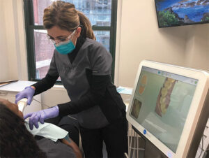
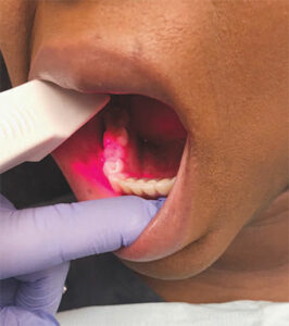
Clinicians with appropriate knowledge and training can complete a full-mouth scan in 3 minutes to 5 minutes.5,16 Patients are immediately shown images of fractured teeth, deteriorating restorations, calculus, and gingival inflammation (Figure 1). Oral health professionals can monitor orthodontic relapse, malocclusion, attrition, abfraction, and any changes in gingival recession.2 Scans can effectively show occlusion and its relevance to oral health. Patients can see areas of heavy occlusal wear and periodontal conditions that may be related to occlusal trauma. Images can also be used to evaluate crowding, overjet, and overbite. Discussing orthodontic treatments to achieve ideal occlusion and prevent trauma is made easier by the use of intraoral scans.
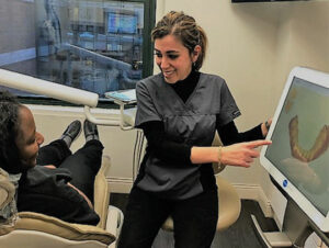
Patients can now view digital replications of their mouths, with alternative visual perspectives (Figure 2). Dental hygienists can focus on areas of concern and establish self-care regimens based on the needs of the patient. The intraoral scan can also be used when the patient returns for follow-up hygiene visits. Clinicians and patients can visually review previous scans, take new scans, and compare changes. Digital acquisition units using 3D imaging enable dental hygienists to show patients oral health conditions in a much more effective way than simple verbalization of dental findings.
DIGITAL IMPRESSIONS
Digital impressions decrease patient discomfort considerably when compared with conventional physical impressions.3,5,16 The digital scan eliminates the need for materials and impression trays, which are often uncomfortable for patients.12,15 According to the literature, patients prefer digital impressions over conventional impressions.4,8,11–13,17 Conventional physical impressions may cause temporary discomfort, as the technique requires the material to be in the mouth until it sets.1,2,5 Patients with strong gag reflexes or excess saliva and children are the least likely to tolerate conventional impression procedures.2,5,10 With digital impressions, If the patient is experiencing discomfort, the scan can be paused and restarted as often as needed.1,5,10,12
Additionally, digital impressions prevent the need to retake impressions, which create more patient discomfort and prolong the appointment.2,5,10,16 Intraoral scans eliminate the need to pour the model in stone in addition to selecting different tray sizes, disinfecting trays, sending impressions to labs, and enduring significant wait times.6 Digital impressions are filed electronically and indefinitely, reducing the space needed for storing impressions and models.18 Intraoral scans can simply be deleted and re-scanned immediately without having to repeat the whole procedure.5,10,16 The images are magnified and errors are captured immediately. Storing the images on the computer also supports efficient record keeping and a paper-free environment.
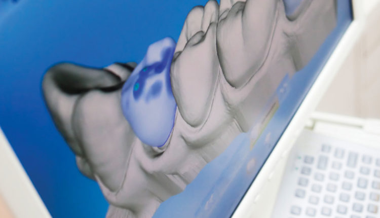
Intraoral scanners differ in accuracy, speed, wand size, wand weight, and image color, depending on the manufacturer.5 The latest generation of scanners has a wider scan range and can be used for full-arch scanning. Older scanners may be limited in scan range and are best for single restorations.5 This is an important aspect to consider when incorporating intraoral scanners into daily clinical use. In addition to the scan range, the scanning speed should be considered. New scanners are generally faster than older ones. However, even with faster scanners available, the speed of the scan depends on the experience level of the clinician. As such, appropriate training is necessary. Improvements in newer generation scanners include the size of the tip and weight of the wand. Tips are now smaller in size, which are easier to use around molars and more comfortable for the patient.5 Lightweight wands also provide ergonomic benefits.5–7 Some of the recent scanners can obtain accurate in-color 3D models of the dental arches being scanned, as opposed to the generic color provided by older scanners. (Figure 3).19 This is especially meaningful when explaining gingival conditions to patients.5
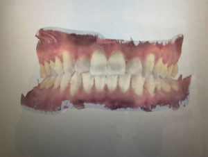
First-generation scanners typically used powder and opacification.5,13 Dental tissues may present with many reflective surfaces, such as enamel crystals, polished surfaces, and amalgam fillings and crowns, which could disrupt the scan.5,13 To prevent this, some intraoral scanners require a powder to be placed on the surface to increase diffuse light. Placing the powder is relatively uncomfortable for patients as it may get contaminated with saliva, which requires removal and reapplication. Applying too much powder may change the thickness of area being scanned and reduce image quality. Applying powder can be time consuming when scanning full arches.13,14 In order to avoid powder, the clinician may change the orientation of the camera to increase diffuse light (Figure 4). Recently introduced intraoral devices can detect optical impressions without the use of powder. Latest-generation scanners have a wider clinical application use, increased diagnostic capabilities, and reduced scanning time.3,5,10
TRAINING REQUIRED
Using the intraoral scanner requires training for all team members. Clinicians with a greater affinity for technology tend to adapt to the new tool more quickly.10 To better understand the technology and its advantages, offices utilizing intraoral scanners will need to cross-train the dental team.10,16
All dental team members need to be comfortable discussing the advantages of the scans used in the office. Knowledgeable clinicians, for example, can describe how CAD/CAM restorations can help retain maximum tooth structure and why this is important to the strength of the tooth and highlight their benefits such as one-visit restorations.1,2,9
COST CONSIDERATIONS
Training is only a portion of the preparation needed to effectively incorporate intraoral scanning technology into a dental practice. Each intraoral scanner has different components that dental offices need to fit into the daily workflow. The initial cost of incorporating intraoral scanning units can be significant. When integrating a digital system into a dental practice, several factors influence cost, including the ability to mill restorations in office.
Intraoral scanners can be purchased in open-format, which allows the clinician to immediately open the file once scanned. This format is preferred not only because it is versatile, but because it also reduces future costs incurred in a closed format, such as the need to purchase specific licenses and pay an annual or monthly fee to unlock files. When purchasing open format intraoral scanning devices, a certain degree of experience and knowledge in computers is needed to integrate the system into the existing office workflow.
Clinicians who lack computer and software integration skills might opt to purchase a closed format. Closed systems are fully proprietary and do not integrate with other components from other companies. Understanding the different technologies available, along with the expectations of the clinician, are important when selecting the proper device.14
CONCLUSION
The use of intraoral scanners enables better patient education, improved esthetic quality, and increased patient comfort. They reduce the chair time required for treatment and produce highly efficient and accurate images. Digital imaging/impressions and CAD/CAM technology are invaluable to improve efficiency and ensure cohesive care.9,16 As the field of dentistry continues to evolve, dental hygienists will need to remain up-to-date on new technologies that can improve patient care and comfort.
REFERENCES
- Nedelcu R, Olsson P, Nyström I, Thor A. Finish line distinctness and accuracy in 7 intraoral scanners versus conventional impression: an in vitro descriptive comparison. BMC Oral Health. 2018;18:1–11.
- Dunlap T, Keller DC, Louis S, Marshall M V, Costerton JW. Clinical dentistry. J Clin Dent. 2011;22:149–158.
- Lee KM. Comparison of two intraoral scanners based on three-dimensional surface analysis. Prog Orthod. 2018;19:6.
- Gan N, Xiong Y, Jiao T. Accuracy of intraoral digital impressions for whole upper jaws, including full dentitions and palatal soft tissues. PLoS One. 2016;11:1–15.
- Mangano F, Gandolfi A, Luongo G, Logozzo S. Intraoral scanners in dentistry: a review of the current literature. BMC Oral Health. 2017; 17:1-11.
- Beheti MJ, Soni UN, Gharat NV, Mahagaonkar P, Khokhani R, Dash S. Intra-oral scanners: a new eye in dentistry. Austin Journal of Orthopedics and Rheumatology. 2015;2(3):1–7.
- Park HR, Park JM, Chun YS, Lee KN, Kim M. Changes in views on digital intraoral scanners among dental hygienists after training in digital impression taking. BMC Oral Health. 2015; 15:1–7.
- Newby EE, Bordas A, Kleber C, et al. Quantification of gingival contour and volume from digital impressions as a novel method for assessing gingival health. Int Dent J. 2011;61(Suppl 3):4–12.
- Stanley M, Paz AG, Miguel I, Coachman C. Fully digital workflow, integrating dental scan, smile design and CAD-CAM: case report. BMC Oral Health. 2018;18:134.
- Culp L, Wong NY, Misch CE. Digital Technology in Implant Dentistry. 2nd ed. Amsterdam: Elsevier Inc; 2014.
- Hickel R, Dasch W, Mehl A KL. CAD/CAM—fillings of the future. Int Dent J. 1997;47:247–258.
- Ender A, Mehl A. Full arch scans: conventional versus digital impressions—an in-vitro study. Int J Comput Dent. 2011;14:11–21.
- Logozzo S, Franceschini G, Kilpela A, Oy D, Governi L.A Comparative analysis of intraoral 3d digital scanners for restorative dentistry. Available at: semanticscholar.org/paper/A-Comparative-Analysis-Of-Intraoral-3d-Digital-For-Logozzo-Franceschini/be25a0e53ed95965863fdcbe6e76b2edbc67896f. Accessed April 17, 2019.
- Richert R, Goujat A, Venet L, et al. Intraoral scanner technologies: a review to make a successful impression. J Healthc Eng. 2017;2017:8427595.
- Löe H. Ural hygiene in the prevention of caries and periodontal disease. Int Dent J. 2000;50:129–139.
- Logozzo S, Kilpelä A, Mäkynen A, Zanetti EM, Franceschini G. Recent advances in dental optics. Part II: experimental tests for a new intraoral scanner. Opt Lasers Eng. 2014;54:187–196.
- Arakida T, Kanazawa M, Iwaki M, Suzuki T, Minakuchi S. Evaluating the influence of ambient light on scanning trueness, precision, and time of intra oral scanner. J Prosthodont Res. 2018;62:324–329.
- Chun J, Tahk J, Chun YS, Park JM, Kim M. Analysis on the accuracy of intraoral scanners: the effects of mandibular anterior interdental space. Appl Sci. 2017;7:719.
- Nedelcu R, Olsson P, Nyström I, Rydén J, Thor A. Accuracy and precision of 3 intraoral scanners and accuracy of conventional impressions: a novel in vivo analysis method. J Dent. 2018;69:110–118.
From Dimensions of Dental Hygiene. May 2019;17(5):40–43.

This is such a useful technology – it is an environmental improvement – impression material not required, no trays, no pouring up plaster models, eliminate the mess, no impressions and models to disinfect, no outgoing lab bags/slips – it’s all electronic. More importantly more comfortable for the patient; a vast improvement for gaggers and children. Images can be sent electronically to the lab, no carbon emissions in the transport, no courier costs, delivered in a key stroke. Huge time saver all round.
As a clinician I love digital impressions, no mixing materials, no trays for delivery, no premature setting while you let the patient finish their story/question. No chance of getting impression material on their clothing or leaving messy finger prints on their face ‘ruining’ their make up. Less time required as there is no mixing/pouring/separating or trimming. Save on travel time to the lab – simple email the images and lab prescription. No lost misplaced models, model gets broken at the lab, they can make a new one from the file.
Time/staff costs are reduced, material cost are reduced. Patients find them more comfortable, neater and quicker. Appointments are shorter. They have so many practical applications – impressions for bruxing guards, whitening, ortho/invisalign, crown and bridge or simple record taking. They might be pricey upfront, but they’ll pay for themselves in no time.