 RUSN/ISTOCK/GETTY IMAGES PLUS; NECHAEV-KON/ISTOCK/GETTY IMAGES PLUS
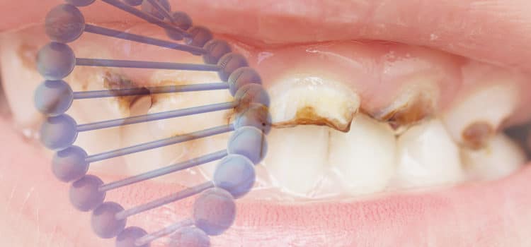
The Role of the Dental Hygienist in Caries Risk Assessment
Dental hygienists are key to personalized, evidence-based caries prevention, which involves a thorough and comprehensive assessment of individual risk indicators and factors, detection and diagnosis of carious lesions, and acknowledgement and incorporation of the patient’s medical and dental histories.
Dental hygienists play a significant role in the assessment of a patient’s individual caries risk. This approach to chronic disease management increases the likelihood that patients will receive appropriate and personalized preventive and therapeutic care.1Modern caries management is determined by risk-based prevention strategies and disease management, centering on identification of the risk and protective indicators, not just the treatment of the disease outcome.2 The process of assessing caries risk should be incorporated into the preventive care visit, while the identification of risk determinants and subsequent risk level designation should be included in the clinical decision-making process for both the clinician and patient.3 Risk assessment recognizes known disease risk factors of dental disease, allowing dental hygienists to effectively aid in promoting personalized preventive strategies specific to that patient’s behaviors and habits—including customizing the frequency of preventive care and recare appointments—and providing oral hygiene education and nutritional counseling.3
CARIES RISK ASSESSMENT
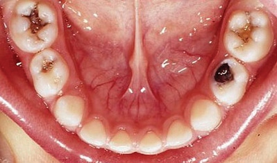
Caries risk assessment is the process of establishing the probability for an individual to develop new enamel or dentin lesions over time (Figure 1).4 The patient is characterized into one of three fixed risk categories: low risk, moderate risk, or high risk.4 Caries risk assessment should be performed on a routine basis, and specifically when life events occur that may influence the balance between de- and re-mineralization of enamel, such as before the eruption of the first and second permanent molars, before the onset of orthodontic appliances, or in the beginning of pregnancy.4 Abanto et al5 found that the use of a caries risk assessment program was effective at reducing the incidence and progression of initial caries lesions in children.5 Afuakwah et al6 reported the use of caries risk assessment improved documentation of caries risk status, leading to improved evidence-based preventive care appropriate to the patient’s risk status.6
CARIES RISK ASSESSMENT MODELS FOR VARIOUS AGES
Various strategies and tools are available for caries risk assessment in daily practice, including an informal assessment, use of structured paper forms, and computer-based programs.4 Separate assessment formats are available for patients ages 0 to 5, as well as patients ages 6 through adult.7–9 Additionally, separate risk assessment protocols for the primary, mixed, and permanent dentitions are available.10,11 Guidelines have been developed based on these various assessment tools to create a framework for categorizing risk level, considerations for treatment options, and determination of individual recare appointments.4
Some examples of caries risk assessment models are the caries risk tool of the American Academy of Pediatric Dentistry, which is used for classifying caries risk in infants, children, and adolescents based on clinical findings and protective and biological factors.3Another model is the American Dental Association caries risk assessment form that evaluates caries risk in infants and children ages 0 to 6 and for patients older than 6.1 Similarly, caries management by risk assessment is an evidence-based caries management philosophy that includes caries risk forms and also protocols that give the clinician management strategies to make restorative, therapeutic, and preventive recommendations for children and adults based on caries risk.7–9 Cariogram is a computer-based caries risk assessment program.12 This is a free downloadable software program designed to calculate the probability of future carious lesions.13 The interaction between 10 caries risk factors is considered, and the caries risk profile of the individual is graphically illustrated.4 The profile is useful for motivational interviewing and creating customized preventive treatment.4
Caries risk assessment tools have been developed for interprofessional care of dental caries in the medical setting. For example, the American Academy of Pediatrics, in conjunction with the federal Maternal and Child Health Bureau, created Oral Health Risk Assessment Training for Pediatricians and Other Health Professionals for pediatric patients.14 This training module provides a brief overview of the guidelines considered for caries risk assessments for children.14 The American Academy of Pediatrics has also developed the Smiles for Life National Oral Health Curriculum, which includes a caries risk assessment module.14
COMPONENTS OF CARIES RISK ASSESSMENT
Dental caries is multifactorial and a chronic disease process.13 The three components of caries risk assessment are risk indicators, risk factors, and protective factors. For a clinician, understanding the components of risk assessment and prognosis is an important facet of clinical decision making.13
RISK INDICATORS
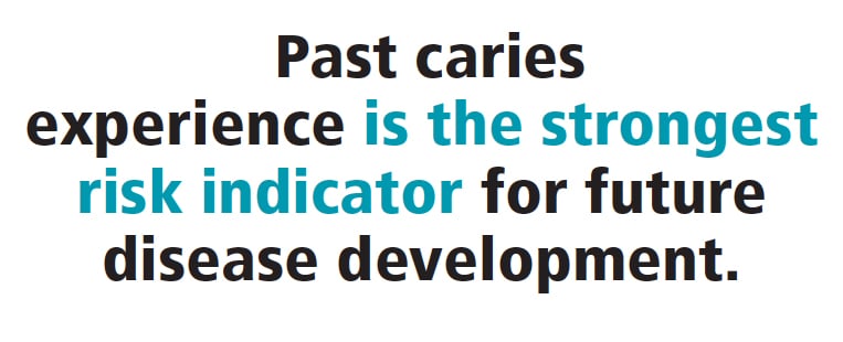
Traditionally, risk indicators associated with dental caries have been defined as clinical observations that illustrate the patient’s history and activity of previous disease development.2 Risk indicators are clinical signs indicating active disease or recent disease.15 These indicators also include variables that are related to disease experience such as socioeconomic status or education.2 Caries experience is an example of a risk indicator that demonstrates how the host copes with biological activity.13
Caries experience. Past caries experience is the strongest risk indicator for future disease development.1 It is a simple, inexpensive, and fast predictor because it only requires a dental examination.2 If interproximal lesions are included in the risk analysis, then radiographs would improve the diagnosis.2 Past caries experience helps to summarize the collective effect of all risk factors and protective factors an individual has been exposed to over a lifetime.2 Also, it is important to include noncavitated lesions in addition to cavitated lesions.2 If decay is present, it is imperative to determine whether it is active or arrested, as this will influence the analysis of future risk.2 Presence of current activity would indicate a high likelihood that if conditions are unchanged, future activity will continue.2
Sociodemographic indicators. Socioeconomic status is a caries risk indicator and predictor in children and adults.16,17 Generally, dental caries is more prevalent in lower than higher socioeconomic classes.2 Research has indicated that children from low-income families have the highest prevalence of early childhood caries.18 In analyzing a patient’s caries risk, the dental hygienist must consider the social environment and lifestyle factors that impact the patient (eg, education, income, occupation, etc).2
RISK FACTORS
A risk factor has an essential role in the etiology of the disease, while a risk indicator is indirectly associated with the disease.2Moreover, caries risk factors are the biologic factors that have caused or contributed to the disease process, or will contribute to its future manifestation on the tooth surface (eg, bacteria, diet, etc).4
Saliva. Saliva maintains an important role in the health of soft and hard tissues in the oral cavity.2 Oral health professionals can assess several salivary parameters related to caries risk, however, the most common ones include salivary flow rate, buffering capacity, and pH level.2 Poor salivary flow rate has been found to be one of the strongest salivary indicators for a high caries risk.19Currently, many medications cause hyposalivation in a percentage of the population and may increase their caries risk.2 Smith et al20revealed dry mouth was the most common side effect of 200 of the most frequently prescribed medications in the United States. Also, certain systemic diseases, especially those related to decreases in salivary flow rate, such as Sjögren syndrome and uncontrolled diabetes, can increase the risk of caries.2 The ability to assess salivary flow chairside provides dental hygienists with the opportunity to identify patients experiencing salivary gland hypofunction and develop personalized treatment options to help remineralization.
Bacteria. Dental caries is caused by the interaction between cariogenic microbial species, such as Streptococcus mutans, and sugary foods and drinks on tooth enamel and/or dentin.21 S. mutans breaks down sugars causing an acidic environment in the plaque, which can result in demineralization of tooth structure over time, leading to dental caries.21 S. mutans is known as an initiator in the caries process, while lactobacilli play a significant role in enhancing the progression of tooth decay.22 In the initial phase, dental caries appears as white spot lesions and progresses to more severe cavitated lesions spreading across the surface of the tooth.21
Diet. Frequent sugar exposure is an important etiologic factor in caries development.2 Dietary considerations include the retentiveness of the food, frequency of consumption, the presence of protective factors in foods (eg, calcium, fluoride), and the type of carbohydrate.2 Dental hygienists developing caries prevention and management treatment options need to recognize the behaviors that are placing the patient at an increased caries risk.23
![]() PROTECTIVE FACTORS
PROTECTIVE FACTORS
 PROTECTIVE FACTORS
 Caries protective factors are biologic or therapeutic factors or measures that can collectively challenge risk indicators or factors that increase caries risk.2 The more prevalent the risk factors, the more protective factors are needed to balance or reverse the demineralization process.2 The most significant protective factor is exposure to fluoride.2 The widespread use of fluoride has helped to reduce the prevalence and rate of progression of dental caries.24 Additionally, the use of fluoride allows for more conservative management strategies for the prevention and treatment of dental caries.2 The application of sealants; the use of strategies to support neutral pH in the oral cavity, such as arginine bicarbonate and calcium carbonate, and remineralization, including calcium phosphate technologies; consumption of oral health products with xylitol; and the use of antimicrobials to control biofilm may also help reduce caries risk.16,25,26
THE ROLE OF DENTAL HYGIENIST
Dental hygienists are the primary preventive specialists of the dental team and are in a unique position to implement office-based caries risk assessment/management programs.1 Through a clinical operations model practiced by most dental offices, dental hygienists have the availability to ensure that a caries risk assessment is completed for every patient, documented properly in the chart, and reassessed routinely at subsequent recare appointments. This information can be used to expand the preventive and clinical care therapies available for caries management based on each patient’s individual needs and risk, as well as aid the clinician in measuring success overtime.1 Effective chronic disease management through caries risk assessment and management can positively impact and reduce caries disease among patients in all age demographics.1 Oral health professionals must recognize that the process of personalized, evidence-based caries prevention and management is multifaceted and involves a thorough and comprehensive assessment of individual risk indicators and factors, detection and diagnosis of carious lesions, and acknowledgement and incorporation of the patient’s medical and dental histories.1
REFERENCES
- Francisco EM, Johnson TL, Freudenthal JJ, et al. Dental hygienists’ knowledge, attitudes and practice behaviors regarding caries risk assessment and management. J Dent Hyg. 2013;87:353–361.
- Fontana M, Gonzalez-Cabezas C. Evaluation du risque carieux chez l’adulte. Réalités Cliniques. 2011;22(3):213–219.
- American Academy of Pediatric Dentistry. Guideline on caries-risk assessment and management for infants, children, and adolescents. Pediatr Dent. 2013;35:E157–E64.
- Twetman S, Fontana M, Featherstone JD. Risk assessment—can we achieve consensus? Community Dent Oral Epidemiol. 2013;41:e64–70.
- Abanto J, Celiberti P, Braga MM, et al. Effectiveness of a preventive program based on caries risk assessment and recall intervals on the incidence and regression of initial caries lesions in children. Int J Paediatr Dent. 2015;25:291–299.
- Afuakwah C, Welbury R. Why do you need to use a caries risk assessment protocol to provide an effective careis preventive regime? Prim Dent J. 2015;4:56–59, 61–66.
- Ramos-Gomez FJ, Crall J, Gansky SA, et al. Caries risk assessment appropriate for the age of 1 visit (infants and toddlers). J Calif Dent Assoc. 2007;35:687–702.
- Featherstone JDB, Domejean-Orliguet S, Jenson L, et al. Caries risk assessment in practice for age 6 through adult. J Calif Dent Assoc. 2007;35:703–713.
- Jenson L, Budenz AW, Featherstone JDB, et al. Clinical protocols for caries management by risk assessment. J Calif Dent Assoc. 2007;35:714–723.
- Evans RW, Pakdaman A, Dennison PJ, et al. The Caries Management System—an evidence-based preventive strategy for dental practitioners. Application for adults. Aust Dent. 2008;53:83–92.
- Evans RW, Dennison PJ. The Caries Management System: an evidence-based preventive strategy for dental practitioners. Application for children and adolescents. Aust Dent J. 2009;54:381–389.
- Bratthall D, H€ansel Petersson G. Cariogram—a multifactorial risk assessment model for a multifactorial disease. Community Dent Oral Epidemiol. 2005;33:256–64.
- Fontana M. The clinical, environmental, and behavioral factors that foster early childhood caries: evidence for caries risk assessment. Pediatr Dent. 2015;37:217–225.
- American Academy of Pediatrics. Oral Health Education and Training. Available at: aap.org/en-us/advocacy-and-policy/aap-health-initiatives/Oral-Health/Pages/Education-and-Training.aspx. Accessed June 26, 2018.
- Featherstone JDB. The caries balance: contributing factors and early detection. J Cal Dent Assoc. 2003;31:129–133.
- National Institutes of Health. Diagnosis and Management of Dental Caries Throughout Life. Available at: consensus.nih.gov/2001/2001DentalCaries115PDF.pdf. Accessed June 26, 2018.
- Jamieson LM, Mejía GC, Slade GD, et al. Predictors of untreated dental decay among 15-34-year-old Australians. Community Dent Oral Epidemiol. 2009;37:27–34.
- Ghazal T, Levy SM, Childers NK, et al. Prevalence and incidence of early childhood caries among African-American children in Alabama. J Public Health Dent. 2015;75:42–48.
- Leone CW, Oppenheim FG. Physical and chemical aspects of saliva as indicators of risk for dental caries in humans. J Dent Educ. 2001;65:1054–1062.
- Smith RG, Burtner AP. Oral side-effects of the most frequently prescribed drugs. Spec Care Dentist. 1994;14:96–102.
- Colak H, Dülgergil CT, Dalli M, et al. Early childhood caries update: a review of causes, diagnoses, and treatments. J Nat Sci Biol Med. 2013;4:29–38.
- Conrads G, About I. Pathophysiology of dental caries. Monogr Oral Sci. 2018;27:1–10.
- Zero D. Sugars–the arch criminal? Caries Res. 2004;38:277–285.
- United States Centers for Disease Control and Prevention. Recommendations for Using Fluoride to Prevent and Control Dental Caries in the United States. Available at: cdc.gov/mmwr/preview/mmwrhtml/rr5014a1.htm. Accessed June 26, 2018.
- Cantore R, Petrou I, Lavender S, et al. In situ clinical effects of new dentifrices containing 1.5% arginine and fluoride on enamel de- and remineralization and plaque metabolism. J Clin Dent. 2013;24(Spec No A):A32–A44.
- Neville BW, Damm DD, Allen CM, Chi AC. Oral and Maxillofacial Pathology. 4th ed. St. Louis: Elsevier; 2016: 912.
From Dimensions of Dental Hygiene. July 2018;16(7):21-24.
