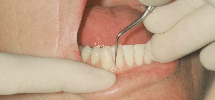
Using Files in Periodontal Therapy
File application in very tenacious calculus removal and overhanging amalgam restorations can be beneficial for both clinician and patient.
Click here to view a file instrumentation assessment form.
Have you wanted to use a file during debridement therapy but were unsure of the technique? Have you experienced tenacious calculus or an amalgam overhang that you could not remove with ultrasonic inserts or curets? File application in periodontal therapy is extremely valuable in certain therapeutic situations.
Although calculus is not an etiology of periodontitis, it is a major contributing factor that permits bacteria to form mature biofilms. Therefore, calculus removal is integral to the success of periodontal therapy. Indications for files include removal of tenacious calculus and overhanging amalgam restorations. Very tenacious calculus can initially form by the build-up of layers over a significant time period. Light deposits are characteristically not well-attached to the surface and are removed with flexible shank curets and ultrasonic instrumentation. However, the mode of attachment of light calculus is sometimes tenacious and fine finishing files are advantageous in this situation. Moderate and heavy calculus can be tenacious and is often more firmly attached than light deposits. Heavy deposits are usually, but not always, tenacious and attach fairly well to tooth surfaces creating a need for ultrasonic instruments or files.
In addition, tenacious calculus can occur when a deposit is smoothed prior to removing its mass.1-3 Inadvertent burnishing can be caused by inappropriate instrument selection and/or technique. If the curet blade-to-tooth angle is too closed (less than 70°) or if the blade is dull, the deposit is smoothed instead of removed. Also burnishing will occur if lateral pressure with a curet is not adequate or if short, powerful channeling strokes are not applied. A smooth deposit is difficult to detect during instrumentation. The intermittent use of an explorer during therapy will aid the clinician in detection. Additionally, scheduling of a 4-to-6 week reevaluation allows for the reassessment of the root surface prior to the maintenance visit.

The addition of perioscopy-the use of the dental endoscope in periodontal therapy-enhances the clinician’s ability to visualize biofilm, root deposits, granulation tissue, caries, and root fractures.4 Perioscopy reveals that as much calculus as possible must be removed because, even after extensive ultrasonic and hand instrumentation, persistent inflammation exists adjacent to residual calculus.1-3 Research studies also conclude that, despite our best efforts, calculus remains on tooth surfaces ranging from 17% to 64% after closed scaling and root planing and 7% to 24% after surgical intervention and open instrumentation by experienced operators.5 File use in therapy will aid in reducing burnished or residual calculus and plaque-retentive overhanging amalgam restorations.
Periodontal files remove the bulk of amalgam overhangs and refine the restorative margin. The files crush and fragment the overhanging ledge prior to removing the restorative material by channeling strokes with a gold knife and/or curet. Files are used for small, medium, or large amalgam overhangs in conjunction with reciprocating handpieces, curets, gold knives, and various armamentarium.
Periodontal files are contraindicated for calculus that is not tenacious and that is removable with other manual or ultrasonic instrumentation. Also, files are contraindicated for overhanging restorations that are a flash or Type I size because a curet, gold knife, and/or a reciprocating handpiece are adequate for removal.
File Design
Each file is composed of a head (body), shank, and handle. The round, oval, oblong or rectangular head contains multiple cutting edges that are adapted parallel to the calculus deposit and engaged, resulting in the fracturing of the calculus. The portion of the cutting edge facing the shank and handle, called the lip, is located at either a 90° or 105° angle with the shank. Rank angle refers to the distance between each lip that approximates 55° depending on the design. Shanks of files are either straight or contra-angled, and vary in length and width to facilitate adaptation.
Files are divided into two classes based on their function-working or finishing (see Figure 1 for examples). Working files have cutting edges that are further apart than files used to finish root surfaces. The working file is usually associated with a large and bulky head; however, the Hirschfeld series have very small heads permitting adaptation subgingivally. The Hirschfeld #3/11 design is for buccal and lingual surfaces, the #5/11 is for mesial and distal surfaces, and the #9/10 file is for the anterior areas. These files can be used on any surface depending on ability to adapt appropriately. The Hirschfeld design houses three cutting edges each of which are sharpened at the first sign of dullness. The Hirschfeld files are extremely useful because of the small head and few cutting edges making them effective and efficacious on tenacious deposit and overhanging amalgam restorations.1,2,6,7 The Orban series are also working files. The #10/11 is for buccal and lingual and the #12/13 is for the mesial and distal surfaces.

Finishing files include the Bedbug UW B that has a medium size head and about 10 cutting edges. Fine files (Bedbug) are used for light deposit, root roughness or grainy areas, and final finishing of overhang removal. A specific application is removing light or moderate deposit located on the cementoenamel junction. For example, finishing files are indicated at a re-evaluation visit if a site did not heal and a discernible calculus deposit is not detectable, but graininess exists on the root surface. Maybe the deposit(s) is quite small and flat, and is mechanically attached, making removal difficult. Applications for finishing files are limited when compared to working files used for moderate to heavy tenacious deposit removal.
Diamond coated files are also available for removal of biofilm, light calculus, and/or residual fine calculus.1,2,8 They are designed for furcations and root depressions for mesial and distal line angles and developmental grooves. The double ended instruments are round or curet-like in design with a 180° or 360° diamond coating to enhance access and versatility. Horizontal, oblique, and vertical stroke patterns are used. Diamond coated files are not designed the same as the other files discussed here. They will be addressed in subsequent articles. Resources for the diamond coated files appear in Table 1.
Instrumentation with files has received considerable criticism because of the potential for creating root surface roughness. This outcome can be avoided if files are used directly on calculus deposits. File use is not indicated in the removal of bacterial plaque or endotoxins because the endotoxin or lipopolysaccharide derived from cell walls of gram negative bacteria is only weakly adherent to periodontally involved root surfaces;9 therefore, it is readily removed with other instruments.
Another criticism of files is their inability to adapt subgingivally to root anatomy due to the bulky design. However, the file is only adapted to the heavy or moderate deposit as other instruments are used to remove the remainder of the calculus adjacent to the cementum. In fact, access with files with very narrow heads is better than curets in some situations. Small working files have a narrower depth diameter than the curet that promotes easy insertion into the periodontal pocket, especially with tight tissue tone.10 For this reason, they are very effective near the junctional epithelium where it is not feasible to insert a curet under the calculus due to lack of space (see Figure 2). The use of files on deposits attached to curved root surfaces, such as line angles or in furcations, is limited because of the flat design of the head containing the cutting edges. Working files are useful for removing tenacious deposits from developmental depressions and grooves.
Recommended Technique
Positioning, grasp, and fulcrum placement are similar to those used with curets and are maintained to adapt the file head parallel to the surface being treated. Adaptation and activation of files differs from curets in blade-to-tooth relationship and stroke activation. A file is placed on top of calculus and not on the root surface. Because it is not always possible to assess calculus location and configuration with a file, the clinician explores the deposit well and mentally visualizes the location and size on the root. The file is then placed to match the visual image and, hopefully, the bulk of a heavy deposit is detectable. The additional use of perioscopy identifies burnished or hard to detect calculus; thus, facilitating the use of files during reevaluation and periodontal maintenance appointments.
With the file parallel to the deposit the cutting edges are engaged by applying pressure on the finger rest, thumb, and index fingers. A long pull stroke is not used because eventually the file’s cutting edges will reach root surface not covered with calculus, causing cemental striations. Instead, the stroke should be very short and directed into the calculus, forcing the deposit into the rake angle resulting in crushing and fragmenting. After each of these strokes, the clinician relaxes the grasp and replaces the file head on the deposit.
To effectively fracture deposits, the file head is adapted parallel to the calculus in various directions. The deposit can be approached with the shank or handle placed vertically, obliquely, or horizontally to facilitate fragmentation. In other words, the crushing stroke is multidirectional to penetrate the mass of deposit from different angles. To do so, a variety of fulcrums are needed. Most frequently, conventional fulcrums are used. Cross arch fulcrums can be employed for proximal surfaces and opposite arch fulcrums for maxillary posterior areas.
Sequencing of instruments in nonsurgical periodontal therapy is critical for success. Periodontal files are used at the beginning of therapy prior to ultrasonic and curet use, or used interchangeably with traditional ultrasonic inserts prior to curet use. In both cases, it is advisable to check the deposit(s) with an explorer after removal to assess if moving on to precision-thin ultrasonic inserts or curets is appropriate. If tenacious deposit still exists, files are again applied until the deposit is reduced to a light deposit that can be removed with finer instruments.
Files are also useful when a patient’s health status contraindicates the use of ultrasonic instrumentation for tenacious deposits. The file takes the place of the ultrasonic entirely and its use is followed by sickles and curets.
Sharpening Technique
The purposes and objectives of sharpening a file are the same as for a curet. Ensuring that each of the multiple cutting edges is sharpened to efficiently engage a calculus deposit or amalgam overhang is important. The rake angle of the file must first be recognized (see Figure 3). The tanged file is then placed within the rake so that the tanged file is positioned toward the handle; thus, the 55° angle portion of the lip is sharpened (see Figure 3). Sharpening the 90° angle (lips parallel to the end of the file head) results in wear in a vertical direction and an increase in the rake and the lip angles. These two effects decrease the file’s longevity and increase the time necessary to achieve sharpness.10 Diamond coated files do not require sharpening.

To sharpen the file, the clinician must ensure the tanged file is parallel to the cutting edge on both sides of the file’s head. The file head is first positioned parallel to a surface (a counter or the floor). The tanged file is then placed with the rake parallel to the file head and surface of the floor. Once positioned, the tanged file is moved back and forth (horizontally) in short strokes across each adjacent cutting edge maintaining the parallel relationship throughout the stroke.
One way to stabilize the sharpening device and instrument is to hold the tanged file in a palm grasp with one hand, grasp the instrument with the head supported on the index finger of the other hand, and tuck arms close to the body for stability. The clinician holds the instrument with no movement and moves only the sharpening device (tanged file) in the back and forth motions that are straight and even.10 The same ideal positioning is achieved by bracing the file against an object (counter) to keep it stationary, ensuring the face is parallel with the floor, and moving the tanged file appropriately.

The final product is not easily evaluated by observing the file edges with a magnifying glass because the “white line” on a beveled area of a curet is not as easily visible on a file. Therefore, the best tests for evaluating sharpness are tactile ones: biting on a test stick and evaluation (gripping) on the deposit. A microscope or magnification loupes are useful to examine the cutting edges. Although sharpening with various devices yields inconsistent results,11 it is still a necessity to enhance effectiveness in therapy. One study demonstrated that new file blades were extremely variable in sharpness, thus suggesting a need to sharpen prior to use as is recommended with curets.11
In summary, periodontal files are used primarily for crushing and fragmenting heavy to moderate hard, tenacious deposits during initial therapy. Periodontal files can be applied to light calculus if its mode of attachment is tenacious. Also, periodontal files are used for amalgam overhang removal. Although the use of files in periodontal instrumentation may be limited, when tenacious deposits and overhanging amalgam restorations exist they are useful in preparing the biologically acceptable root surface.
References
- Pattison AM, Matsuda S. Making the right choice. Dimensions of Dental Hygiene. 2003;1(8)(Suppl):4-10.
- Matsuda S. Instrumentation of biofilm. Dimensions of Dental Hygiene. 2003;1(1):26-30.
- Pattison AM, Pattison GL. Periodontal instrumentation transformed. Dimensions of Dental Hygiene. 2003;1(2):18-22.
- Stambaugh RV. Perioscopy-the new paradigm. Dimensions of Dental Hygiene. 2003;1(2):12-16.
- Drisko CL, Killoy WJ. Scaling and root planing: removal of calculus and subgingival organisms. Current Opinion in Dentistry. 1991;1:74-80.
- Nield-Gehrig JS. Fundamentals of Periodontal Instrumentation and Advanced Root Instrumentation. Philadelphia: Lippincott Williams and Wilkins; 2004:361-373.
- Hodges, KO. Instrument selection: philosophy and strategies. In: Hodges KO, ed. Concepts in Nonsurgical Periodontal Therapy. Albany, NY: Delmar Publishers; 1998:276-277.
- Duff BC. Duff Diamond Furcation Files. Periodontics Online. 1999. Available at: http://www.periodontics/online.com/duffdiamond files.html. Accessed July 24, 2004.
- Kieser JB. Nonsurgical periodontal therapy. In: NP Lang, T Karring, ed. Proceedings of the 1st European Workshop on Periodontology. London: Quintessence Publishing Co Ltd; 1993.
- Hoople S. Files provide desirable results in patient treatment procedures. RDH. 1985:22-24.
- Pasquini R, Clark SM, Baradaran S, Adams DF. Periodontal files-a comparative study. J Periodontol. 1995;66:1040-1046.
From Dimensions of Dental Hygiene. November 2004;2(11):16, 18-20.

