Tips for Identifying Systemic Conditions and Anomalies on the Panoramic Image
Dental professionals can leverage panoramic imaging to uncover systemic conditions, improving both oral care and overall health outcomes.
This course was published in the January/February 2025 issue and expires February 2028. The authors have no commercial conflicts of interest to disclose. This 2 credit hour self-study activity is electronically mediated.
AGD Subject Code: 730
EDUCATIONAL OBJECTIVES
After reading this course, the participant should be able to:
- Discuss the importance of panoramic images in identifying systemic conditions and anomalies.
- Describe the radiographic features seen in various conditions.
- Identify how to avoid errors when capturing panoramic images.
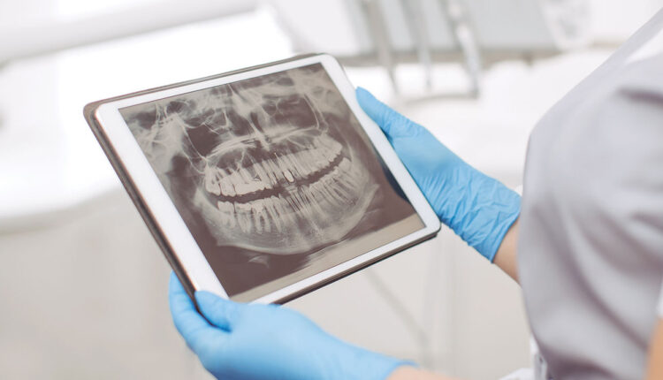
In the realm of medical diagnostics, the panoramic image has emerged as a valuable tool. It provides a large field of view to assess growth and development and identify the presence of third molars, trauma, as well as other findings. The panoramic image can also reveal unexpected systemic issues, shedding light on broader health concerns. As dental hygienists are trained in the interpretation of radiographic images, the importance of meticulous inspection during interpretation cannot be overemphasized.
Osteoporosis
Osteoporosis is a progressive bone disease characterized by decreased bone density and strength, leading to an increased risk of fractures.1 Most commonly impacting older adults, it can also occur in all populations and age groups.1,2 An estimated 10 million Americans age 50 and older have osteoporosis, some unknowingly, due to its asymptomatic progression.1–3 Osteoporosis can lead to pain in the lower back, pathological fractures, loss of height, and severe kyphosis.1–3
 A density scan, or bone densitometry, of the lumbar spine and hips is used to diagnose osteoporosis.4 However, bone pattern changes may sometimes be identified by using the mandibular cortical thickness on a panoramic image.1,5,6 Classification is determined by measuring the width of the cortex distally from the mental foramen, and assigning a mandibular cortical index (Figures 1-4).5
 Dental professionals may be able to identify changes in the cortical plate and refer patients to their physician. In addition, dental hygienists can encourage lifestyle modifications and educate patients regarding the risk of osteonecrosis, a rare complication of bisphosphonate therapy.

Benign Lesions
Cemento-osseous dysplasia presents with changes in bone patterns in which normal bone is replaced with benign, fibro-osseous lesions. Over time, tissue calcifies with osseous and cementum-like tissue.7 This condition is divided into three subgroups: focal, periapical, and florid. The focal group is typically located around mandibular molars, the periapical group is often noted around the apex of mandibular anterior teeth, and florid lesions may be seen in both the maxillary and mandibular arches.7-10 In most cases, these lesions are asymptomatic and are an incidental finding during periodic radiographic examinations. Complications may arise if an individual has failed endodontic work in the area of a cemento-osseous dysplasia. The lesion could become symptomatic at this point and require surgery. 7-10
Odontomas may also be identified on a panoramic image.10 These lesions are a grouping of benign tumors that consist of epithelium and primitive connective tissue called ectomesenchyme. The etiology is not always known, but is thought to arise from developmental abnormalities. Rarely symptomatic, odontomas may be identified by dental practitioners after radiographic examinations.
An odontoma can be either compound or complex in nature. Compound odontomas are typically located in the incisor cuspid region of the maxilla and can have a more “tooth-like” appearance. Complex odontomas are likely found in the premolar molar region of the mandible, and present more as a disorganized mass of tissue. Odontomas are more likely to be associated with adult dentition and are rarely observed in primary dentitions.10-12 Figure 5A and B show a mixed radiopaque/radiolucent, multilocular lesion that begins mid-root between the mandibular left premolars and extends beyond the apices. Panoramic imaging is particularly suited for observing these lesions. Due to the advancement in imaging, cone-beam computed tomography (CBCT) scans can now provide improved diagnostic information about these lesions.7,8,11,12
 Cemento-osseous dysplasia and odontomas are mostly asymptomatic and may not require any intervention. Dental hygienists who observe a lesion on a periapical film, CBCT, or panoramic image play an important role in initiating the referral process. Patients may be advised to remove odontomas to eliminate the concern of cystic changes, bone destruction, or interference with unerupted teeth. It is important to note the various ways in which a lesion may present and periodically update images of asymptomatic lesions that are being watched.7,9,10
Calcification of the Carotid Arteries
When reviewing a panoramic image, dental professionals should consider bony and soft tissue structures.13 The dental hygienist is able to observe areas of the carotid artery as they appear on a panoramic image. The bifurcated area of the carotid artery may become calcified, narrowing the vessel. Atherosclerosis occurs when an artery narrows from plaque or calcification.14-16 Calcification of the carotid artery may be noted by the appearance of a radiopaque area near the vertebra. Often, this opacity will be visible lateral and inferior to the hyoid bone, and may be unilateral or bilateral (Figure 6).15,17
 Images may require adjustment by toggling the brightness and contrast to identify calcification. A study by Janiszewska-Olszowska et al17 found evidence of calcification in 21% of panoramic images reviewed. If carotid artery calcification is identified, the patient should be referred to his or her primary care physician for further evaluation.
Oculo-Facio-Cardio-Dental Syndrome
While rare and only impacting women, oculo-facio-cardio-dental (OFCD) syndrome also has cardiovascular implication that may be identifiable on a panoramic image..18,19 Patients with OFCD are often identified in the dental setting due to abnormally long canine roots (radiculomegaly) viewable in panoramic images (Figure 7).19,20 Patients with OFCD often have canines that continue to develop until they reach the cortical plate of the orbit.21 Oral health professionals may also notice facial elongation, a high nasal bridge, and a broad nasal tip. However, as the name denotes, OFCD also causes congenital cataracts and congenital heart diseases. The cause of OFCD has been identified as a genetic x-linked dominant disorder.18–22
 OFCD is diagnosed by identifying the abnormally long canines, which may appear as if they are extending into the orbit of the eye, in a panoramic image. Once this is noted, the clinician may see other abnormalities such as the facial features mentioned above or crowding, clefts, delayed eruption, or missing teeth.18-20 Patients with these presentations should be referred for genetic testing.
Orthodontia may be difficult for individuals with OFCD due to the long roots of the canines. Other prostheses, such as implants or bridges, may be required to replace missing teeth. Oral hygiene instruction should emphasize good self-care to prevent endodontic therapy, which may be challenging.18,20,22
Eagle Syndrome
Also rare, eagle syndrome may be detected on a panoramic image. The underlying cause of eagle syndrome has not been determined; however, theories range from trauma leading to reactive hyperplasia to the expansion of osseous tissue at the origin of the stylomandibular ligament. Facial and neck pain are common symptoms of eagle syndrome. Eagle syndrome is diagnosed by identifying clinical symptoms, a physical exam, and palpitations of the tonsillar region. Administration of lidocaine into the anterior pillar and the tonsillar fossa may also be used to aid in diagnosis and identification of where the pain is originating from.23-26
CT scans are generally considered the gold standard for diagnostic imagery of eagle syndrome due to their ability to demonstrate the relationship between the bony styloid process, the nerve, and vascular anatomy. However, a panoramic image can also be useful in diagnosing eagle syndrome, or at the very least lead to a referral for further investigation.24
Nonsurgical treatment includes pharmacotherapies such as anti-analgesics and acetaminophen. Additionally, antiseizure and antidepressants have been prescribed as well as gabapentin. Those who do not wish to undergo surgery may choose to receive regular injections of local anesthetics and corticosteroids. Unfortunately, the therapeutic effects of the injections lessen over time.24-26 A more long-lasting and definitive treatment is surgery.24,25
Avoiding Errors
Due to the wide view provided by the panoramic image, oral health professionals are able to visualize the sinuses, temporomandibular joint, and hyoid bone.13,27 When considering conditions that could present on a panoramic image, producing a high-quality, diagnostic image is critical. To achieve this, the dental hygienist must focus on the patient position and patient instruction before exposure.13,28
Positioning errors are common and may impact diagnosis, leading to repeat radiation exposure.13,27-29 The most common patient positioning error is incorrect placement of the tongue. If the patient’s tongue is not positioned against the palate for the duration of image acquisition, a radiolucent shadow covers the apices of the maxillary anterior teeth, making it difficult to diagnose the area.13,27 Other common positioning errors include a slumped spine, resulting in a radiopaque spinal column covering the lower center of the image; the chin tipped either too high or too low, resulting in an image with an exaggerated frown or smile; the patient placed too far forward or too far back on the bite stick, causing anterior teeth that are too skinny or wide; the patient’s midsagittal plane not centered, leading to a disproportionate ramus; and a ghost image artifact from metal that was not removed prior to exposure.31,13,27-30
The patient may move during the process, which causes a ripple or blurring of the image. These errors can be avoided when clinicians ensure their patient is positioned correctly and understands what to do.13,27-30 The clinician may want to demonstrate the necessary positioning.27,28 Emphasizing the importance of keeping the tongue in place during entire exposure is important. Careful instruction should be given to hold still, stand up straight, hold onto the machine handles, and take a step forward.
The dental hygienist should watch to ensure the patient is adhering to these instructions.13,27,28 Critical positioning and removal of all metal from the neck up should be methodically approached like a checklist to ensure the clinician has not overlooked any aspect of proper alignment.13,31 By devising a systematic process for panoramic image exposure, the dental hygienist’s efforts are more likely to result in an image that not only shows typical orofacial structure, but also any anomalies that may be present.27–31
Conclusion
At some point, dental hygienists will come across unusual findings during radiographic evaluations. Staying abreast of the various ways systemic conditions and other anomalies could present on a panoramic image is integral to optimal patient-centered care. Maintaining keen eyes to distinguish normal anatomy from abnormal findings is key to exceptional dental hygiene care.
References
- Mupparapu M., Akintoye SO. An application of panoramic radiography in the detection of osteopenia and osteoporosis – current state of the art. Curr Osteoporos Rep. 2023; 21:354-359.
- Friedlander AH, Norman KM, Farman AG, Nortje CJ, Wood RE. Panoramic Radiographic Detection of Systemic Disease. Panoramic Radiology. 2007:167-182.
- National Institute on Aging. Osteoporosis. Available at nia.nih.gov/health/osteoporosis/osteoporosis. Accessed December 5, 2024.
- Radiological Society of North America. Bone Density Scan (DEXA or DXA). Available at radiologyinfo.org/en/info/dexa. Accessed December 5, 2024.
- Shintaku WH, Encisco R, Covington JS, Migliorati CA. Can dental students be taught to use dental radiographs for osteoporosis screening? J Dent Edu. 2013;77:598-603.
- Sukegawa S, Fujimura A, Taguchi A, et al. Identification of osteoporosis using ensemble deep learning model with panoramic radiographs and clinical covariates. Sci Rep. 2022;12:6088.
- Ravikumar S, Vasupradha G, Menaka T, Sankar S. Focal cemento-osseous dysplasia. J Oral Max Path. 2020;24:19-22.
- Tidke P, Gupta N, Patil D, et al. Periapical cemento-osseous dysplasia: A journey from diagnostic dilemma to accurate diagnosis with use of 3D imaging. J Pharm Bioallied Sci. 2024;16:951-S954.
- Prodromidis GI, Tosios KI, Koutlas IG. Cemento-osseous dysplasia-like lesion and complex odontoma associated with an impacted third molar. Head Neck Pathol. 2011;5: 401–404.
- Borghesi A, Tonni I, Pezzotti S, Maroldi R. Peripheral osteoma, compound odontoma, focal cemento-osseous dysplasia, and cemento-ossifying fibroma in the same hemimandible: CBCT findings of an unusual case. J Radiol Case Rep. 2017;12:756-759.
- Sharma R, Prabhadevi MC. Odontome: A brief overview. Int J Clin Pediatr Dent. 2011;4:177-185.
- Khalifa C, Omami M, Garma M, Slim A, Sioud S, Selmi J. Compound‐complex odontoma: A rare case report. Clin Case Rep. 2022;10:e05658.
- Iannucci JM, Howerton LJ. Dental Radiology Principles and Techniques. 6th ed. St Louis, Missouri: Elsevier Inc; 2022.
- Agacayak KS, Guler R, Karatas SP. Relation between the incidence of carotid artery calcification and systemic diseases. Clin Interv Aging. 2020;3:821–826.
- Kwon YE, An CH, Choi KS, An SY. Comparison of carotid artery calcification between stroke and nonstroke patients using CT angiographic and panoramic images. Dento Maxillo Facial Radiology. 2022;51:20210500.
- National Library of Medicine. Carotid Artery Disease. Available at medlineplus.gov/carotidarterydisease.html. Accessed December 4, 2024.
- Janiszewska-Olszowska J, Jakubowska A, Gieruszczak E, Jakubowski K, Wawrzyniak P, Grocholewicz K. Carotid artery calcifications on panoramic radiographs. Int J Environ Res Public Health. 2022;19(21):14056.
- Kato J, Kushima K, Kushima F. New radiological findings and radiculomegaly in oculofaciocardiodental syndrome with a novel BCOR mutation: A case report. Medicine. 2018;97:e13444.
- Nguyen TT, Truong ATH, Hoang VA, et al. Oculo-facio-cardio-dental (OFCD) syndrome: a case report. J Med Case Rep. 2024;18:18.
- Smith MH, Cohen DM, Bhattacharyya I, Islam NM, Kashtwari D. Radiculomegaly: a case report of this rare dental finding with review of the associated oculo-facio-cardio-dental syndrome. Oral Surg Oral Med Oral Pathol Oral Radiol. 2018;126:e220-e227.
- Oh SH, Kang JH, Kang JH, et al. Radiculomegaly of canines in oculofaciocardiodental syndrome. Oral Radiol. 2019;35:326-330.
- Sharifi A, Kouhi A. Management of eagle syndrome. Curr Opin Otolaryngol Head Neck Surg. 2023;31:276-280.
- Dudde F, Bergmann W, Schuck O, Schunk J, Barbarewicz F. Accuracy in the diagnostics of styloid process – panoramic radiograph vs. computed tomography: a comparative study. In Vivo. 2024;38:1390-1396.
- Badhey A, Jategaonkar A, Anglin Kovacs AJ, et al. Eagle syndrome: A comprehensive review. Clin Neurol Neurosurg. 2017; 159:34-38.
- Pagano S, Ricciuti V, Mancini F, et al. Eagle syndrome: an updated review. Surg Neurol Int. 2023;14:389.
- de Barros JF, Rodrigues MV, Barroso LA, Amado IC. Eagle syndrome: an underdiagnosed cause of orofacial pain. BMJ Case Reports. 2021;14:e238161.
- Lingam AS, Koppolu P, Abdulsalam R, Reddy RL, Anwarullah A, Koppolu D. Assessment of common errors and subjective quality of digital panoramic radiographs in a dental institution, Riyadh. Ann Afr Med. 2023;22:49-54.
- Subbulakshmi A, Mohan N, Thiruneervannan R, Naveen S, Gokulraj S. Positioning errors in digital panoramic radiographs: A study. J Orofac Sci. 2016;8:22-26.
- Perschbacher S. Interpretation of panoramic radiographs. Aust Dent J. 2012;57:40-45.
- Alsufyani N, Alnamlah S, Mutaieb S, et al. Virtual reality simulation of panoramic radiographic anatomy for dental students. J Dent Educ. 2023; 87:1200-1209.
- Kim JW, Seo YS. An atypical case involving real, ghost, and pseudo-ghost images on a panoramic radiograph. Imaging Sci Dent. 2024;54:57-62.
From Dimensions of Dental Hygiene. January/February 2025; 23(1):36-41.
