The Dynamics of Pressure
Implementing the correct force for the task at hand will help clinicians improve outcomes and reduce injury.
Controlled application of force in the use of hand instrumentation is an important element of nonsurgical periodontal therapy. This force is produced when pressure is applied by the hand and fingers during instrument activation. Controlled application of pressure affects the efficiency and efficacy of instrumentation, as well as operator ergonomics. Understanding pressure dynamics can help clinicians achieve the best outcomes while safeguarding their musculoskeletal health. During instrumentation, force is applied in three ways: pinch pressure from the grasp fingers, stabilizing pressure of the fulcrum and/or hand rest, and lateral pressure against the tooth prior to and during stroke activation. Each application uses a level of pressure specific to the task. For example, the pressures exerted during exploratory strokes are different from those used with calculus removal strokes. Errors in grasp, fulcrum, and lateral pressures can contribute to ineffective instrumentation.
GRASP PRESSURE AND ERGONOMICS
The upper extremity musculoskeletal disorders most frequently experienced by dental hygienists are often caused by repetitive strain and cumulative trauma from high pinch forces of the grasp used in periodontal instrumentation.1–3 Inexperienced clinicians tend to exert consistently heavy pressure as they learn to control the instrument,4,5 but this can become a debilitating habit.6 In order to prevent injury, clinicians must be cognizant of the pressure used in their grasp.
Grasp pressure on the instrument handle changes according to the task at hand. Insertion, placement, and assessment strokes call for light pressure in order to maximize tactile sensitivity as the instrument tip or blade travels across the tooth or root surface. Surface vibrations transmitted to sensory nerves of the fingertips are heightened when an even, delicate pressure is used. This same light pressure is implemented to confirm the soft tissue attachment—whether probing, exploring, or preparing for a working stroke with a bladed instrument.
Working strokes require an increase in grasp pressure that should vary in intensity based on a deposit’s bulk, degree of mineralization, and tenacity. The pressure exerted from the three grasp points—index finger, middle finger, and thumb—should also vary, as opposed to the even application used in a light-pressured assessment grasp. A working stroke requires force to be directed laterally toward the surface being instrumented. Once the placement stroke is made (light pressure) and soft tissue attachment confirmed, lateral pressure is applied prior to and during activation, then released at completion. The process is repeated for subsequent strokes: light pressure for placement, firm lateral pressure for the working stroke, followed by release.
FULCRUM PRESSURE FOR STABILIZATION
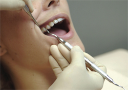
The type of fulcrum used, which provides a stabilizing point for the instrument, changes based on what area is being instrumented and the type of stroke needed. A fulcrum may be an intraoral finger rest or an extraoral hand rest, requiring light, moderate, or firm pressure. For example, extraoral fulcrums are preferred for the maxillary posterior region to facilitate proper angulation of the instrument and to help the operator maintain a neutral wrist position (Figure 1).7 In terms of pressure, probing calls for light-pressured fulcrums whereas calculus removal requires firm pressure on the fulcrum—whether extraoral or intraoral.8
LATERAL PRESSURE AND BURNISHING
The goal of nonsurgical periodontal therapy is to produce a clean, hard root surface free of contaminants.9,10 Partially scaled, burnished deposits create a microscopic biofilm niche that sustains infection in the soft tissue overlying the root.11 Burnishing occurs readily when instruments are dull, but even sharp instruments can burnish calculus on a root surface when insufficient lateral pressure is applied to the blade. When inadequate lateral pressure is used, small increments of deposit are shaved from its outermost surface, leaving the base layer of the calculus intact—smooth and burnished rather than cleanly removed. This may be one of the most common errors in periodontal therapy. Returning maintenance patients who present with bleeding points invariably have burnished calculus on these surfaces, as evidenced by endoscopic studies.12
ANALYTICAL APPROACH TO INSTRUMENTATION
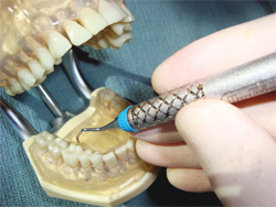
Grasp pressure and lateral pressure integrate in dynamic fluctuation during activation. The lateral pressure used to engage the blade against the deposit is regulated by the grasp and originates from the digits facing the surface to be treated, ie, the thumb, middle finger, index finger, combination of thumb and index finger, or index finger and middle finger, depending on the tooth surface being treated (Figure 2 and Figure 3). Using this model prevents the three-point “death grip” grasp that contributes to musculoskeletal injury.
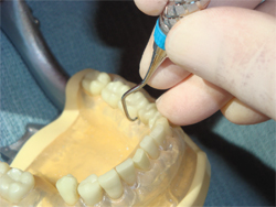
In order to maintain the correct adaptation, angulation, and pressure needed for a precisely controlled working stroke, while also protecting musculoskeletal health, clinicians must determine the appropriate body, arm, and wrist position; fulcrum; and grasp for the task at hand. To accomplish this, clinicians must first visualize the instrument’s position against the tooth surface to determine which digits will provide the vector of force necessary for a good working stroke. This should then determine the position and fulcrum used.
BALANCING PRESSURES FOR SUCCESS
The relationship between the pressure used in the grasp and the fulcrum is often overlooked. A light-pressured grasp should be supported with a light-pressured fulcrum or hand rest. A moderate grasp with lateral pressure for biofilm debridement calls for a moderately-secure fulcrum pressure to stabilize the working stroke. Mineralized deposit removal requires strong lateral pressure for activation, and an equally strong fulcrum pressure (Figure 1).
Adequate lateral pressure must be used with calculus removal strokes, which need to be supported by equivalent pressure into the fulcrum—whether an intraoral finger rest or extraoral hand rest is used.8 In other words, the lateral force directed into the tooth surface by the blade prior to activation must equal the force exerted by the fulcrum finger on a tooth or by the hand against the patient’s extraoral soft tissue.8 Clinicians may be reluctant to apply firm extraoral hand rest pressure against the face of a patient. Without sufficient fulcrum force, however, the extraoral hand rest will lack the stability needed to control a working stroke for calculus removal. A well-controlled instrument is preferred to an unstable one that is slipping off the tooth surface. Stroke control on mineralized deposits is facilitated by lateral pressure that equals fulcrum pressure. The failure to balance these pressures will result in loss of stroke control.
Incorporating pressure dynamics into instrumentation helps clinicians improve instrumentation control and therapy outcomes, while also reducing the risk of injury. The correct application of force in both grasp and fulcrums takes careful consideration, but the extra time spent yields significant benefits for both clinicians and patients.
REFERENCES
- Villanueva A, Dong H, Rempel D. A biomechanical analysis of applied pinch force during periodontal scaling. J Biomech. 2007;40:1910–1905.
- Al Qattan MM, Bowen V. Cumulative trauma disorders of the hand and wrist. Current Opinion in Orthopaedics.1993;4(4):68–71.
- Shenkar O, Mann J, Shevach A, Ever-Hadani P, Weiss P. Prevalence and risk factors of upper extremity cumulative trauma disorder in dental hygienists. Work. 1998; 1:263–275.
- Dong H, Loomer P, Villanueva A, Rempel D. Pinch forces and instrument tip forces during periodontal scaling. J Periodontol. 2007;78:97–103.
- Radwin RG, Oh S, Jensen TR, Webster JG. External finger forces in submaximal five-finger static pinch apprehension. Ergonomics. 1992;35:275–288.
- Lalumandier JA, McPhee SD. Prevalence and risk factorsof hand problems and carpal tunnel syndrome among dental hygienists. J Dent Hyg. 2001;75:130–134.
- Meador HL. The biocentric technique: a guide to avoiding occupational pain. J Dent Hyg. 1993;67:38–51.
- Pattison A, Matsuda S, Pattison G. Extraoral fulcrums. Dimensions of Dental Hygiene. 2004;2(10):20–23.
- Cobb CM. Non-surgical pocket therapy: mechanical. Ann Periodontol. 1996;1:443–490.
- American Academy of Periodontology. Guidelines for the management of patients with periodontal diseases. J Periodontol. 2006;77:1607–1611.
- Matsuda S. Anatomy of a stroke. Dimensions of Dental Hygiene. 2008;6(11):22–26.
- Checchi L, Montevecchi M, Checchi V, Zappulla F. The relationship between bleeding on probing and subgingival deposits: an endoscopical evaluation. Open Dent J.2009;28:154–160.
From Dimensions of Dental Hygiene. September 2012; 10(9): 36; 38-39.
