Strategies for Reducing Staining After Silver Diamine Fluoride Treatment
The use of potassium iodide, potassium fluoride, silver nitrate, and glass ionomer cement may mitigate the discoloration caused by SDF.
The increased prevalence of dental caries among adults over the past few decades has led to new solutions to address this issue. According to the World Health Organization,1 caries is a global healthcare issue that affects an estimated 2 billion people around the world. Adults may be at increased risk of caries due to the presence of gingival recession leading to root exposure and poor oral hygiene.
Treating caries also comes with a high cost, representing about 10% to 15% of global health expenditures every year.1 Surgical and restorative approaches have long been used to treat dental caries, but these processes are expensive and highly invasive. Alternative approaches using minimally invasive procedures have since been suggested.2–5
Use of Silver Diamine Fluoride
The most recent approach to treating dental caries is silver diamine fluoride (SDF). A topical solution, SDF promotes remineralization and hardens carious lesions.6,7 It is designed to arrest root caries, prevent recurrent caries, and reduce sensitivity.8 See the sidebar for how SDF is applied.9
SDF works through the synergistic effect of its silver and fluoride ions, which not only foster alkalinity, an ideal pH for teeth to flourish, but also arrest the processes that contribute to decay.8,10 For instance, SDF inhibits the breakdown of exposed collagen matrix, which provides the scaffold needed for remineralization.8
Surendranath et al8 list five modes of action for SDF:
- Obturates dentinal tubules and stops bacterial invasion
- Penetrates dentin and increases mineral content of the tooth through interaction with hydroxyapatite crystals
- Provides antimicrobial properties
- Inhibits matrix metalloproteinases and halts caries process
- Alkaline properties help to form fluorhydroxyapatite by reacting with calcium and phosphates in saliva
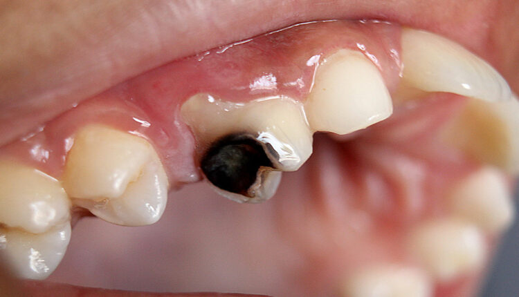
SDF has gained popularity due to its effectiveness in arresting caries through a noninvasive and cost-effective approach.11,12 However, the potential of SDF to stain teeth after treatment has presented an esthetic issue that tends to limit its use despite its high level of clinical efficacy.
This issue has sparked research into various mitigative strategies, including the post-application of potassium iodide (KI), use of potassium fluoride (KF) and silver nitrate (AgNO3), and administration of a glass ionomer cement (GIC). Thus far, the evidence demonstrating the effectiveness of these approaches is limited.
Potassium Iodide Post-Application
KI has been recommended in myriad settings as a remedy for reducing the tooth staining that arises from the use of SDF to arrest dental caries.3,5 It works by reacting with SDF residue to form the transparent compound, silver iodide (AgI).5 AgI is highly photosensitive and dissociates into silver and iodine when exposed to light, eradicating the staining effect left by SDF treatment.5
A systematic review critically evaluated 11 studies of SDF modifications to mitigate staining and concluded that majority of studies showed tooth staining reduction when KI was applied the same day as SDF.2 On the other hand, a systematic review by Roberts et al3 found that staining resurfaced 7 to 14 days after treatment with SDF and KI. Future research should focus on how to make the anti-staining effects initiated by KI more permanent.5
While there is not enough evidence to prove KI’s ability to reduce discoloration, Aly and Yousry5 established that the immediate application of KI after SDF application reduced discoloration. An in vivo study found that the use of a KI solution decreased the amount of discoloration caused by SDF on demineralized dentin.13 The use of SDF and KI together can also enhance remineralization of the dentin.13
KI’s ability to reduce SDF’s black staining is dose-dependent. Detsomboonrat et al14 demonstrated that the use of 20% KI provided the best results in reducing discoloration, but further experiments must be conducted to establish the optimal dose.14
Potassium Fluoride and Silver Nitrate
Little research has been done on the effectiveness of KF and AgNO3 in eliminating SDF-induced staining.2 KF is a well-established agent for caries prevention, known for its lack of impact on enamel color and the absence of white opacity lesions associated with demineralization. When used in conjunction with AgNO3 following SDF application, it yields intriguing results.
The antimicrobial properties of silver complement the process, enhancing remineralization.2 This suggests that the concurrent application of KF and AgNO3 is not an effective solution for preventing SDF staining but could serve as an alternative to SDF, particularly in children.
Glass Ionomer Cement
The latest and most effective strategy to reduce staining caused by SDF is the use of a glass ionomer cement (GIC) restoration after SDF application.13 GIC is a self-adhesive product commonly used in dental practice for restorative purposes. It is supplied as a powder-liquid complex made of fluoro-aluminosilicate glass and an aqueous solution of polyacrylic acid.15
The thixotropic nature of the liquid is seen in its ability to alter thickness on shaking or warming. GIC has been recommended for restorative purposes due to its ease of placement and marginal adaptation.16 As such, following SDF treatment, the use of GIC can help prevent staining and boost longevity of the treatment.
Alsagob et al17 explored whether applying a composite restoration, in comparison to a GIC restoration, following SDF treatment would lead to reduced discoloration. Additionally, they examined whether there was a difference in discoloration when restoration application was immediate or delayed. Results of the in vivo study found that postponing the restoration until 2 weeks after SDF application resulted in a decrease in staining.
In some clinical settings, a GIC restoration is done immediately following the application of SDF. However, this practice is not recommended as research shows the immediate use of GIC upon SDF application may hinder GIC’s ability to reduce staining.2
On the other hand, when GIC is used between 1 and 2 weeks following SDF application, the restorative features of the cement also significantly reduce tooth discoloration.16,19,20 Zhao et al13 observed marginal staining around GIC restorations between 7 and 14 days after treatment.
The effectiveness of GIC post-SDF application is also affected by its ability to adhere to the treated tooth. Adhesive capabilities are measured using the variable shear bond strength (SBS).13 Studies show that SDF use affects the binding strength of GIC in atraumatic restorative treatment.13,18
GIC adhesion uses both chemical bonding mechanisms and microchemical interlocking processes. Restoration using GIC forms optimal SBS when done at least 1 week after SDF treatment. Additionally, the restorative process using GIC is not affected by the immediate application of KI to counter discoloration. The SBS for GIC remains optimal even with the use of KI after SDF.13
Conclusion
Evidence shows that the use of SDF to arrest caries is an effective strategy. However, esthetic concerns hinder the wide adoption of SDF. Mitigative procedures have been suggested using different processes. Thus far it seems the most ideal strategy for reducing SDF-mediated staining is through the application of KI immediately after the process, followed by a GIC restoration process after at least 1 week to maintain optimal SBS.
Further in vivo investigations are needed to better understand the variables present in the biophysiological environment of the oral cavity. Additionally, more evidence is needed on how to instill a longer-lasting effect of KI against SDF-mediated dentinal staining.
References
- World Health Association. Oral Health. Available at: https://www.who.int/news-room/fact-sheets/detail/oralhealth#:~:text=Globally%2C%20an%20estimated%202%20billion,from%20caries%20of%20primary%20teeth. Accessed August 22, 2023.
- Asghar M, Omar RA, Yahya R, Yap AU, Shaikh MS. Approaches to minimize tooth staining associated with silver diamine fluoride: a systematic review. J Esthet Restor Dent. 2023;35:322-332.
- Roberts A, Bradley J, Merkley S, Pachal T, Gopal JV, Sharma D. Does potassium iodide application following silver diamine fluoride reduce staining of tooth? A systematic review. Aust Dent J. 2020;65:109-117.
- Yengopal V, Harneker SY, Patel N, Siegfried N. Dental fillings for the treatment of caries in the primary dentition. Cochrane Database Syst Rev. 2009;2:CD004483.
- Aly MM, Yousry YM. Potential discolouration of silver diamine fluoride versus silver diamine fluoride/potassium iodide in primary teeth: a randomised clinical study. Br Dent J. 2022 Dec 6;1-6.
- Mei ML, Nudelman F, Marzec B, et al. Formation of fluorohydroxyapatite with silver diamine fluoride. J Dent Res. 2017;96:1122-1128.
- Chen J , Yu Z , Zhu P , et al. Effects of fluorine on the structure of fluorohydroxyapatite: a study by XRD, solid-state NMR and Raman spectroscopy. J Mater Chem B. 2015;3:34-38.
- Surendranath P, Krishnappa S, Srinath S. Silver diamine fluoride in preventing caries: a review of current trends. Int J Clin Pediatr Dent. 2022;15(Suppl 2):S247-S251.
- Stefanou LB. Managing high-risk patients With SDF. Dimensions of Dental Hygiene. 2022;20(7):22-24.
- Mei ML, Nudelman F, Marzec B, et al. Formation of fluorohydroxyapatite with silver diamine fluoride. J Dent Res. 2017;96:1122-1128.
- Abdellatif HM, Ali AM, Baghdady SI, ElKateb MA. Caries arrest effectiveness of silver diamine fluoride compared to alternative restorative technique: randomized clinical trial. Eur Arch Paediatr Dent. 2021;22:575-585.
- Nguyen TM, Tonmukayakul U, Hall M, Calache H. Cost-effectiveness analysis of silver diamine fluoride to divert dental general anaesthesia compared to standard care. Aust Dent J. 2022;67:352-361.
- Zhao IS, Chu S, Yu OY, Mei ML, Chu CH, Lo ECM. Effect of silver diamine fluoride and potassium iodide on shear bond strength of glass ionomer cements to caries-affected dentine. Int Dent J. 2019;69:341-347.
- Detsomboonrat P, Thongmak P, Lertpayab P, Aiemsri W, Sooampon S. Optimal concentration of potassium iodide to reduce the black staining of silver diamine fluoride. J Dent Sci. 2022;17:300-307.
- Sidhu SK, Nicholson JW. A Review of glass-ionomer cements for clinical dentistry. J Funct Biomater. 2016;7:16.
- Berg JH, Croll TP. Glass ionomer restorative cement systems: an update. Pediatr Dent. 2015;37:116-124.
- Alsagob E, Sawan N, Aladhyan S, Alsalem N, Alshami A, Albluwi S. Silver diamine fluoride with delayed restoration reduces tooth discoloration. Saudi J Biol Sci. 2022;29:1434-1438.
- Ng E, Saini S, Schulze KA, Horst J, Le T, Habelitz S. Shear bond strength of glass ionomer cement to silver diamine fluoride-treated artificial dentinal caries. Pediatr Dent. 2020;42:221-225.
How to Apply Silver Diamine Fluoride
By Lisa B. Stefanou, RDH, MPH
 The clinical application of silver diamine fluoride (SDF) on facial, lingual, and occlusal surfaces is simple.1,2 Protect the lips with petroleum jelly or lip balm. Dental hygienists may prefer scented lip balm to mask the slight smell of ammonia.
- Isolate the tooth with cotton rolls.
- Clean the lesion of food debris with a microbrush or cotton pellet and then dry it.
- Paint SDF onto the clean lesion for 1 minute and allow to air dry.
- While post-operative rinsing with the air-water syringe after SDF application is not necessary, fluoride varnish should be applied to the treated tooth and the remainder of the dentition. The flavor of varnish is appealing and it keeps the SDF attached to the tooth surface. Fluoride varnish also remineralizes the rest of the dentition. It would be safe to assume that if a patient has one area of decay, other areas of demineralization may be present.
5 . Evaluate the lesion for color change and hardness.
For proximal caries mesial and distal surfaces only:3
- Isolate the tooth with cotton rolls.
- Clean the lesion of food debris with floss.
- Place an SDF-coated soft pick into the proximal area for 1 minute and allow to air dry. The pick can be pulled gently in and out to agitate. The SDF then becomes absorbed into the proximal lesion.
- Dab additional SDF using a microbrush above the contact and in the buccal and lingual sluiceways. Blot any excess fluid and blood with a cotton roll, pellet, or swab. If there is radiographic evidence of a deeper decalcification or caries lesion, apply an additional 60-second insertion in the same manner.
- Paint fluoride varnish (5%) over the treatment area with the pick still in place and then withdraw the pick.
- Using radiography, note any changes in lesion 6-months post-application.
- Apply fluoride varnish to the remainder of the dentition.
Lisa B. Stefanou, RDH, MPH, is a clinical associate professor and director of Dental Hygiene Programs at New York University (NYU) College of Dentistry in New York City. She is also the co-developer of NYU College of Dentistry’s accelerated degree track in the dental hygiene program, which provides a pathway for internationally educated dentists to become licensed dental hygienists.
References
- Use of silver diamine fluoride for dental caries management in children and adolescents, including those with special health care needs. Pediatr Dent. 2017;39:146–155.
- Seifo N, Robertson M, MacLean J, et al. The use of silver diamine fluoride (SDF) in dental practice. Br Dent J. 2020;228:75–81.
- Croll TP, Berg J. Delivery methods of silver diamine fluoride to contacting proximal tooth surfaces and history of silver in dentistry. Compend Contin Educ Dent. 2020;41:84–89.
From Dimensions in Dental Hygiene. September 2023; 21(8):14-17.
