Reduce Caries Risk
Effective sealant placement is key to preventing decay in pits and fissures.
Dental hygienists play a crucial role in the prevention of dental diseases, including the selection, preparation, and placement of sealants. United States Centers for Disease Control and Prevention (CDC) data indicate that about 90% of carious lesions are found in the pits and fissures of permanent teeth, with molars being the most susceptible.1,2 The first step in an effective caries-prevention protocol is a thorough caries risk assessment. Assessing risk for disease development is an important component of any disease prevention program. Risk susceptibility can be determined on a variety of levels, including community, individual, tooth, and tooth surface. Since dental caries is a bacteria-dependent, multifactorial disease, preventive measures, such as sealants, can be implemented once the most significant risk factors are identified.3 All types of patients—from an 8-year-old child with newly erupted permanent teeth to a cancer patient undergoing head and neck radiation—can benefit from a customized caries risk assessment.
SEALANT PLACEMENT
Sealants must remain in place and completely cover pits and fissures to be effective. The two factors most likely to affect sealant retention are proper application and the tooth’s eruption status.4,5 While sealant placement is fairly uncomplicated, manufacturer instructions must be followed.
Sealants placed early in eruption are far more likely to need replacement. A study by Dennison et al reported that when an operculum existed over the distal marginal ridge of molars, the sealant replacement rate was 54%.6 In contrast, the replacement rate was 0% for a selected sample of sealants placed at later eruption stages over a 5-year period.7 This creates a dilemma for the practitioner because some permanent molars erupt with fissures that seem at risk of decay. Since they appear at-risk early in the eruption stage, the clinician may opt to seal such surfaces, knowing that replacement may be inevitable.
SEALANT MATERIALS
Pit and fissure sealants today are composed of unfilled and filled resins and are available with fluoride-releasing or glass ionomer cements.8 Sealants are also available in a number of forms, including self-etching, wet/dry field, and colored sealants. Glass ionomer sealants can be applied in very moist conditions and in places where isolation is difficult. They have the ability to uptake and release fluoride, which may increase the ability of fissures to resist demineralization even after sealant material has deteriorated.6,9 Glass ionomer-based sealants generally release higher fluoride amounts than traditional materials.10 The retention of glass ionomer sealants, however, is not as good as traditional materials.11
The latest innovation in sealants is the use of nanotechnology in the development of dental composites. The newly available nanomaterials, such as nanofillers and nanohybrids, can improve the hardness of sealants and ease of application.12 The addition of nanoparticles to resin composites may create flowable materials with both higher mechanical properties and better flow characteristics than traditional materials.13 Ku?göz et al found that nanofilled resin-based fissure sealants showed improved surface hardness, in addition to similar or better fissure sealing ability in compared to other materials tested.14
TOOTH PREPARATION
Different methods of preparing an enamel surface prior to sealant application have been studied, but no one technique has proven superior. As long as the enamel surface is well-cleaned, retention of the sealant should be optimal.15
The conventional technique begins with a pumice prophylaxis, rinsing, drying, and acid etching with 37% phosphoric acid for 30 seconds to 40 seconds. Previous research shows that prophylaxis with pumice and a pointed bristle brush or rubber cup causes greater microleakage.16 It may be that a rubber cup or pointed bristle brush with pumice does not adequately clean pits and fissures so the etchant can produce a surface area receptive for bonding.
The enameloplasty sealant technique (EST) excavates the pits and fissures using a round tungsten carbide bur under low speed. The area is then acid etched for 30 seconds to 40 seconds with 37% phosphoric acid, rinsed, and dried. The main aim of EST is to remove debris and acquired pellicle, open up the fissures, and increase the surface area. An increased surface area enhances sealant retention, thus decreasing microleakage. EST is able to decrease microleakage most likely because it enlarges the narrow fissures, allowing the sealant to easily penetrate and eliminate the acquired pellicle, thus increasing the sealant adaptability.17
The fissurotomy technique uses a microshort tapered fissured bur to open up the fissures. Pits and fissures are prepared to the size of the bur head, followed by acid etching for 40 seconds with a 37% phosphoric acid, rinsing, and drying. Chaitra found that the fissurotomy technique caused the same amount of microleakage as conventional sealant technique, but significantly more than EST.17
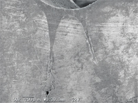
Air abrasion involves using alumina particles to open the pits and fissures with subsequent acid etching. A 2001 study revealed that microleakage can be prevented most effectively with a combination of mechanical air abrasion and chemical acid etching. Air abrasion is not as effective as acid etching in surface roughening, but when used in addition to acid etching, the technique further reduces microleakage. Future studies, possibly using scanning electron microscopy, to investigate the sealant–enamel interface, as well as resin tag formation using air abrasion, are needed.18
Some level of microleakage occurs with all techniques, due to the flowable composite exhibiting some amount of polymerization shrinkage after light curing, which may create microgaps between the tooth and the composite.17
The use of an Er:YAG laser in conjunction with acid etching is the most recent surface preparation technique used. The Er:YAG laser beam (k = 2940 nm) is absorbed by the intrinsic water in the enamel apatite. This leads to the generation of heat and water vapor, which causes micro-explosion and tissue removal. Khogli et al found that both Er:YAG laser and bur enameloplasty methods demonstrated no significant difference when compared to the conventional, noninvasive sealing technique.19 In contrast, previous studies reported enhanced pit and fissures sealant penetration when the bur enameloplasty was used.18
In a recent systematic review of controlled clinical trials that compared different surface cleaning methods directly (surfaces cleaned with a handpiece and prophylaxis brush with pumice, compared to surfaces cleaned only by running an explorer along the fissures and cleaning with an air-water syringe) found no difference in sealant retention.20
TOPICAL FLUORIDE APPLICATION
In clinical practice, patients often receive prophylaxis and fluoride treatment prior to examination by the dentist. In cases where a partially missing sealant is found after the fluoride treatment has been applied, a small touch-up sealant application may be necessary. It was thought that recently-applied fluoride application could render the enamel somewhat more resistant to an adequate acid etching procedure. A study done by Koh et al, however, concluded that topical fluoride has no clinical effect on the retention of pit and fissure sealants.21
RESOURCES
A recent report released by the American Dental Association Council on Scientific Affairs provides an invaluable literature review on pit and fissure sealants published prior to 2008. Published in the Journal of the American Dental Association, this report provides a wealth of information related to the clinical success of pit and fissure sealants.11
The ADA’s expert panel addressed four clinical questions:
- Under what circumstances should sealants be placed to prevent caries?
- Does placing sealants over early (noncavitated) lesions prevent progression of the lesion?
- Are there conditions that favor the placement of resin-based sealants vs glass-ionomer cement sealants in terms of retention or caries prevention?
- Are there any techniques that could improve sealants’ retention and effectiveness in caries prevention?
These questions should serve as an initial assessment and are important clues to the end result of sealant application. Sealants are an important tool in the caries prevention armamentarium, and dental hygienists, as the leaders in preventive dentistry, need to be well-versed on the latest research available on this technique.
REFERENCES
- Centers for Disease Control and Prevention. National Health and Nutritional Examination Survey. Questionnaires, datasets, and related documentation. Available at: www.cdc.gov/nchs/data/nhanes/ survey_content_99_10.pdf. Accessed May 14, 2012.
- Macek MD, Beltrán-Aguilar ED, Lockwood SA, Malvitz DM. Updated comparison of the caries susceptibility of various morphological types of permanent teeth. J Public Health Dent. 2003;63:174–182.
- Rethman, J. Trends in Preventive Care: Caries Risk Assessment and Indications for Sealants. J Am Dent Asso. 2000;131:85–125.
- National Institutes of Health consensus development conference statement on dental sealants and the prevention of tooth decay. J Am Dent Assoc. 1984;108:233–236.
- Simonsen R. Pit and fissure sealant. J Pract Hyg. 1996;1:37–38.
- Markovic DLJ, Petrovic BB, Peric TO. Fluoride content and recharge ability of five glass ionomer dental materials. BMC Oral Health.2008;8:21.
- Dennison JB, Straffon LH, More FG. Evaluating tooth eruption on sealant efficacy. J Am Dent Assoc. 1990;121:610–614.
- Ripa L, Leske G, Varma A. Longitudinal studies of the caries susceptibility of occlusal and proximal surfaces of first permanent molars. J Public Health Dent. 1988;48:8–13.
- Marks, D. Effect of adhesive agent and fissure morphology on the in vitro microleakage and penetrability of pit and fissure sealants. Quintessence Int. 2009;40:763–772.
- Lobo MM, Pecharki GD, Tengan C, da Silva DD, daTagliaferro EP, Napimoga MH. Fluoride releasing capacity and cariostatic effect provided by sealants. J Oral Sci. 2005;47:35–41.
- Beauchamp J, Caufield PW, Crall JJ, et al. Evidence-based clinical recommendations for the use of pit and fissure sealants: a report of the American Dental Association Council on Scientific Affairs. J Am Dent Assoc. 2008;139:257-268.
- Mitra SB, Dong WU, Holmes BN. An application of nanotechnology in advanced dental materials. J Am Dent Assoc. 2003;134:1382–1390.
- Beun S, Bailly C, Devaux J, Leloup G. Rheological properties of flowable resin composites and pit and fissure sealants. Dent Mater. 2008;24:548–555.
- Ku?göz A, Tüzüner T, Ülker M, Kemer B, Saray O. Conversion degree, microhardness, microleakage and fluoride release of different fissure sealants. J Mech Behav Biomed Mater. 2010;3:594–599.
- Simonsen RJ, Neal RC. A review of the clinical application and performance of pit and fissure sealants. Aust Dent J. 2011;56;45–58.
- Chan DC, Summitt JB, García-Godoy F, Hilton TJ, Chung KH. Evaluation of different methods for cleaning and preparing occlusal fissures. Oper Dent. 1999;24:331–336.
- Chaitra TR, Subba Reddy VV, Devarasa GM, Ravishankar TL. Flowable resin used as a sealant in molars using conventional, enameloplasty and fissurotomy techniques: an in vitro study. J Indian Soc Pedod Prev Dent. 2010;28:145-150.
- Hatibovic-Kofman S, Butler SA, Sadek H. Microleakage of three sealants following conventional, bur, and air-abrasion preparation of pits and fissures. Int J Paediatr Dent. 2001;11:409–416.
- Khogli AE, Cauwels R, Vercruysse C, Verbeeck R, Martens L. Microleakage and penetration of a hydrophilic sealant and a conventional resin-based sealant as a function of preparation technique: a laboratory study. Int J Paediatr Dent. 2012 Jan 25. Epub ahead of print.
- Muller-Bolla M, Lupi-Pégurier L, Tardieu C, Velly AM, Antomarchi C. Retention of resin-based pit and fissure sealants: a systematic review. Community Dent Oral Epidemiol. 2006;34:321–336.
- Koh SH, Huo YY, Powers JM, Chan JT. Topical fluoride treatment has no clinical effect on retention of pit and fissure sealants. J Gt Houst Dent Soc. 1995;67:16–18.
From Dimensions of Dental Hygiene. June 2012; 10(6): 36, 38, 41.
