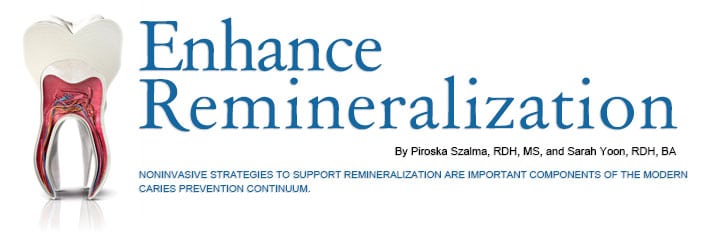
Enhance Remineralization
Noninvasive strategies to support remineralization are important components of the modern caries prevention continuum.
In recent years, the provision of dental services and oral health status have substantially improved. Despite these efforts, 10% to 20% of the United States population still experiences dental caries, and recent epidemiological surveys show that the incidence of dental caries is increasing.1
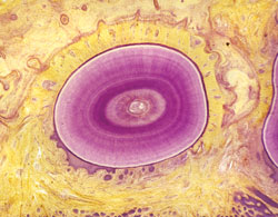
Modern day dietary habits present challenges in maintaining the health of many biochemical properties in the oral cavity. Repetitive exposures to fermentable carbohydrates and acid-rich foods can throw off the balance between the remineralization and demineralization process. Softening of the tooth surfaces—enamel, dentin, and exposed cementum—may also result, increasing the risk of caries lesions (Figure 1).2 Acidogenic bacteria are present in the oral cavity along with other microflora, and their levels are kept in balance through various interactions among the different species.3 When food is ingested, the acid-producing bacteria normally present in the mouth metabolize the available carbohydrates and turn them into lactic acid, effectively lowering the pH of the oral cavity, creating an environment conducive to tooth demineralization.3
Because dental caries is such a dynamic disease process involving the cyclic demineralization and remineralization of tooth structure, maintaining an oral environment favorable to remineralization is key.4 A state of health exists when net mineral loss (demineralization) and net mineral gain (remineralization) remain in equilibrium.5 Although tooth enamel has a high mineral content consisting of 96% calcium hydroxyapatite, it is still vulnerable to demineralization.6 Damaging mechanical and chemical etiologies, such as dental caries, abrasion, abfraction, attrition, and acid erosion, challenge the limited repair ability of the oral cavity on a daily basis.7 By encouraging the process of remineralization, the enamel can compensate for such disadvantages and is able to replace the lost mineral with a less soluble apatite.6
Initial demineralization of enamel begins below the surface, with the outer layers of the enamel remaining intact during early stages of caries development. At this point, the tooth may benefit from therapies that promote remineralization—resulting in an arrest or even a reversal of the caries progression.4
BIOLOGICAL DEFENSE
The main biological defense against demineralization is saliva. Its acid buffering ability, as well as its mineral components, play a significant role in maintaining the balance of remineralization and demineralization.6 The oral cavity is constantly challenged with the change of pH, and the mineral components in saliva—calcium, phosphate, and fluoride ions—assist in remineralizing the tooth structure.6
Another key factor in maintaining a healthy environment in the oral cavity is the acquired enamel pellicle. Predominantly composed of salivary proteins, carbohydrates, nonsalivary proteins, and lipids, the acquired enamel pellicle is a thin, acellular film that forms over tooth surfaces when exposed to the oral environment.8 When the pellicle forms over the tooth structures, it provides a significant defense and resistance to acid challenges and enamel demineralization by preventing direct contact between the acids and the enamel.3,6
Xerostomic or hyposalivary conditions in the oral cavity create an imbalance between demineralization and remineralization. Saliva remineralizes demineralized tooth enamel by providing bioavailable calcium and phosphate ions to the tooth.9 When hyposalivary conditions are present, the reparative ability of saliva is hindered, and it cannot provide organic and mineral material to fill in microscopic defects of the tooth structure. The formation of the acquired enamel pellicle is also slowed, limiting its ability to create a protective barrier against acidic conditions. Therefore, promoting normal salivary flow rate is necessary to prevent demineralization.10
XYLITOL
The addition of xylitol, a naturally occurring sweetener, into the diet may enhance salivary flow and reduce the number of acidogenic bacteria present in the oral cavity.3 Some studies show that when xylitol (at least 4 g to 7 g) is used periodically (three to seven times throughout the day), especially in gum, it can reduce the available fermentable sugars in the mouth while promoting salivary flow, effectively reducing the number of acid-producing bacteria that can lower the pH and demineralize teeth.3 Many health organizations worldwide support the habitual use of sucrose-free xylitol or other sugar alcohol combinations in chewing gum or lozenges for at-risk populations.11 Research is still ongoing to evaluate the efficacy of xylitol in other modalities, such as dentifrices.12
FLUORIDE
Fluoride is very effective in promoting remineralization and is available in several delivery methods with varying levels of concentration and administration times.13 Fluoride can be applied topically via toothpastes, gels, varnishes, foam, and mouthrinses.6
While fluoride is an evidence-based treatment that successfully reduces dental caries, more adjunct therapies, in addition to health promotion methods, are being explored to enhance fluoride’s role in the remineralization of enamel and other tooth surfaces.13 Comprehensive caries prevention protocols should include fluoride and other agents that help create balance between demineralization and remineralization of the tooth structure.13 Fluoride’s ability to promote remineralization is limited by the availability of calcium and phosphate ions present in the saliva.
When fluoride is applied, the saliva needs to contain adequate calcium and phosphate ions to produce remineralization.14 For every two fluoride ions, 10 calcium ions and six phosphate ions are required to form one unit cell of fluorapatite.15 Remineralization occurs when the fluoride, calcium, and phosphate enter the subsurface region of the lesion and form a new layer on the existing crystalline remnants in the lesion.16
CALCIUM PHOSPHATE
The clinical use of calcium and phosphate ions for remineralization was not very successful in the past due to the low solubility of calcium phosphates, especially in the presence of fluoride ions.17 Insoluble calcium phosphates required acid to produce ions that were able to diffuse into the enamel; soluble calcium phosphates did not substantially incorporate into dental plaque or localize at the tooth surface to produce effective concentration to drive the mineral diffusion into the enamel.17 Today, new methods have improved the ability to deliver calcium and phosphate molecules.
Currently, there are four commercially available calcium phosphate-based remineralization systems on the market: amorphous calcium phosphate (ACP); casein phosphopeptide (CPP)-ACP (Recaldent); calcium sodium phosphosilicate (Novamin); and tricalcium phosphate (TCP). ACP delivers a calcium salt and a phosphate salt separately (eg, from a dual chamber device) intraorally. As the salts mix with saliva, they dissolve, releasing calcium and phosphate ions to produce an ion activity product that results in an immediate precipitation of ACP, or in the presence of fluoride ions, amorphous calcium fluoride phosphate.15 In the intraoral environment, these two phases are very unstable and quickly transform to a thermodynamically stable, insoluble crystalline phase such as fluorhydroxyapatite.
This quick transformation of the ions, however, can limit ACP’s ability to keep the ions transiently bioavailable to promote enamel subsurface lesion remineralization.15 In Recaldent, the CPP stabilizes high concentrations of calcium and phosphate ions, together with fluoride ions, at the tooth surface by binding to the acquired salivary pellicle and plaque.18 CPP-ACP increases the number of calcium and phosphate ions that help remineralize the enamel.3 Some studies have shown that it increases calcium and phosphate ion levels in supragingival plaque, promotes the remineralization of enamel subsurface, slows progression of coronal caries, and provides better protection against an acidic environment.6,15,19
Novamin technology is based on a calcium sodium phosphosilicate bioactive glass, which is designed to release calcium and phosphate ions intraorally to help remineralization of teeth.15 Most often used to treat dentinal hypersensitivity, Novamin may also be effective in remineralizing caries lesions and preventing demineralization.15 TCP is a new addition to the calcium phosphate technology market. In TCP, a “functionalized” calcium and a “free” phosphate are created. Because the functionalized calcium remains protected, calcium ions are prevented from interacting with fluoride ions before reaching the tooth surface. Once TCP is exposed to saliva, the calcium is free to release calcium, phosphate, and fluoride ions onto the tooth surface.20
Calcium phosphate technologies are promising treatment modalities and may be beneficial for a variety of patients at increased caries risk, however, more evidence is needed to support their use. The American Dental Association (ADA) Council on Scientific Affairs (CSA) published a systematic review in 2011 that investigated nine studies on the use of calcium phosphate technologies to promote remineralization. Because of the variances in study design, the review stated that the current body of evidence is insufficient to support the use of calcium phosphate technologies to reduce caries.12,21
ANTIMICROBIAL THERAPY
Researchers are also exploring the effectiveness of antimicrobials in reducing the number of acid-producing bacteria in the oral cavity. Chlorhexidine is effective in reducing the number of Streptococcus mutans and other acidogenic and cariogenic bacteria in the mouth.3 However, research shows that chlorhexidine’s effect is short lived. Also, it is not clear if reducing the number of S. mutans alone will decrease decay because other factors affect caries risk, such as xerostomia, diet, and personal oral hygiene.3 Chlorhexidine varnishes may have a longer effect in maintaining a lower number of acidogenic bacteria, but there isn’t enough research to support more widespread use.3
The ADA CSA panel determined that chlorhexidine varnish had a lack of effect on caries incidence in children, although it did find that chlorhexidine/thymol varnish was effective in reducing root caries among adults.12,21
LASERS
Using laser irradiation (eg, Er:Yag laser), in conjunction with fluoride treatment, to promote remineralization has been explored in laboratory settings.22 Some studies have shown that lasers can reduce enamel permeability and inhibit demineralization.23,24 When a low-energy Er:YAG laser is irradiated onto a tooth surface following fluoride treatment, it may inhibit enamel demineralization by a photothermal purification of enamel hydroxyapatite, inhibit enamel diffusion, and deposit fluoride more effectively on the enamel surface, therefore promoting formation of fluoridated hydroxyapatite.22 This would reduce enamel solubility and may help prevent enamel demineralization.22 Study on lasers and remineralization is still in its infancy. Currently, these studies are only conducted in vitro due to the instantaneous temperature rise on the enamel surface, as well as in the tooth pulp, upon laser irradiation. Measuring such temperature rise on tooth structures needs to be investigated further before clinical applications of laser irradiation treatment can be possible.22
CONCLUSION
The demineralization and remineralization cycle is an ongoing process that continuously challenges the balance in the oral cavity.25 Even in a healthy oral environment, minerals are lost and regained because of tooth enamel’s porous structure.26 A hyposalivary oral environment further tips the scale toward demineralization. The goal of modern dentistry is to manage noncavitated caries lesions noninvasively through remineralization in an attempt to prevent disease progression and improve esthetics, strength, and function.8
Remineralization therapy using fluoride and adjunctive treatments can help preserve healthy tooth structure and is less costly than restorative procedures.4 While new therapies are promising, the research remains limited, and more in vivo studies must be conducted in order to demonstrate efficacy.13,21 In order for noninvasive remineralization therapy to be successful, patients must be educated on their use and agree to comply with the suggested treatment plan.25 Patient preference must also be considered, as it plays an important role in determining which products and procedures can be easily incorporated into a person’s daily self-care routine.3 Dental professionals must remain vigilant in their monitoring of caries activity and the success of implemented therapies.4,6
ACKNOWLEDGEMENT
CLAUS LUNAU / SCIENCE SOURCE IMAGES
ASTRID AND HANNS-FRIEDER MICHLER/SCIENCE SOURCE
REFERENCES
- Bagramian RA, Garcia-Godoy F, Volpe AR. The global increase in dental caries: a pending public health crisis. Am J Dent. 2009;22:3–8
- Cooper B. Protecting your healthy patients.Dimensions of Dental Hygiene. 2008;6(4):38–39.
- Su N, Marek CL, Ching V, Grushka M. Caries prevention for patients with dry mouth. J Can Dent Assoc. 2011;77:b85.
- Diefenderfer KE, Stahl J. Caries remineralization therapy: implications for dental readiness. Mil Med. 2008;173(Suppl):48–50.
- Silverstone LM. Remineralization phenomena.Caries Res. 1977;11(Suppl):59–84.
- Willis, S Hays, R. Partner with your patients:how to keep the remineralization/demineralization process in balance. Dimensions of Dental Hygiene. 2011;9(3):58–63.
- Øgaard B. The cariostatic mechanism of fluoride. Compend Contin Educ Dent. 1999;20(Suppl):10–17.
- Siqueira WL, Custodio W, McDonald EE. New insights into the composition and functions of the acquired enamel pellicle. J Dent Res. 2012;91:1110–1118.
- Cochrane NJ, Cai F, Huq NL, Burrow MF,Reynolds EC. New approaches to enhanced remineralization of tooth enamel. J Dent Res. 2010;89:1187–1197.Amaechi BT, Higham SM. In vitro remineralization of eroded enamel lesions by saliva. J Dent. 2001;29:371–376.
- Fontana M, González-Cabezas C. Are we ready for definitive clinical guidelines on xylitol/polyol use? Adv Dent Res. 2012;24:123–128.
- Pickett FA, Tornwall R. Nonfluoride caries prevention. Dimensions of Dental Hygiene. 2012;10(12):40–43
- ten Cate JM. Novel anticaries and remineralizing agents: prospects for the future. J Dent Res. 2012;91:813–815.
- Fujikawa H, Matsuyama A, Uchiyama S,Nakashima T, Ujiie T. Influence of salivary macromolecules and fluoride on enamel lesion remineralization in vitro. Caries Res.2008;42:37–45.
- Reynolds EC. Calcium phosphate-based remineralization systems: scientific evidence? Aust Dent J. 2008;53:268–273.
- ten Cate JM, Featherstone JD. Mechanistic aspects of the interactions between fluoride and dental enamel. Crit Rev Oral Biol Med.1991;2:283–296.
- Gurunathan D, Somasundaram S, Kumar SA.Casein phosphopeptide-amorphous calcium phosphate: a remineralizing agent of enamel. Aust Dent J. 2012;57:404–408.
- Reynolds EC. Casein phosphopeptide amorphous calcium phosphate: the scientific evidence. Adv Dent Res. 2009;21:25–29.
- Reynolds EC, Cai F, Shen P, Walker GD.Retention in plaque and remineralization of enamel lesions by various forms of calcium in a mouthrinse or sugar-free chewing gum. J DentRes. 2003;82:206–211.
- Karlinsey RL, Mackey AC, Walker ER, RederickKE. Preparation, characterization, and in vitro efficacy of an acid-modified beta-TCP material for dental hard-tissue remineralization. Acta Biomater. 2010;6:369–978.
- American Dental Association Council onScientific Affairs. Non-fluoride caries preventive agents: full report of a systematic review and evidence-based recommendations. Available at:ebd.ada.org/contentdocs/clinical_recommendations_non_fluoride_caries_preventive_agents_full_report.pdf. Accessed April 22, 2013.
- Liu Y, Hsu CYS, Teo CMJ, Teoh SH. Potential mechanism for the laser-fluoride effect on enamel demineralization. J Dent Res. 2013;92:71–75.
- Maung NL, Wohland T, Hsu CY. Enamel diffusion modulated by Er:YAG laser (Part 1)—FRAP. J Dent. 2007;35:787–793
- 4. Fried D, Featherstone J, Visuri S, Seka W, Walsh J. The caries inhibition potential of Er:YAG and Er:YSGG laser radiation. SPIE Proceedings. 1996;2672:73–78.
- Cochrane NJ, Cai F, Huq NL, Burrow MF,Reynolds EC. New approaches to enhanced remineralization of tooth enamel. J Dent Res. 2010;89:1187–1197.
- Gugnani N, Gupta M, Josan R, Pandit I. Cariesinfiltration of noncavitated white spot lesions: a novel approach for immediate esthetic improvement. Contemp Clin Dent. 2012;3(Suppl):199–202.
- Hegde MN, Devadiga D, Jemsily PA.Comparative evaluation of effect of acidic beverage on enamel surface pre-treated with various remineralizing agents: An in vitro study. J Conserv Dent. 2012;15:351–356.
-
From Dimensions of Dental Hygiene. May 2013; 11(5): 42, 44, 46.
