 GILAXIA/ ISTOCK/GETTY IMAGES PLUS
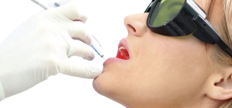
The Use Of Lasers in Nonsurgical Periodontal Therapy
The efficacy of this technology as an adjunctive treatment for periodontitis and peri-implant diseases remains controversial.
This course was published in the February 2017 issue and expires February 2020. The author has no commercial conflicts of interest to disclose. This 2 credit hour self-study activity is electronically mediated.
EDUCATIONAL OBJECTIVES
After reading this course, the participant should be able to:
- List the uses for each laser technology in periodontal therapy.
- Discuss the evidence regarding laser use as an adjunct to scaling and root planing.
- Describe the effectiveness of photodynamic therapy used for antimicrobial purposes in periodontal treatment.
- Explain the ethical and legal considerations related to dental hygienists’ use of lasers.
Although lasers have been used in periodontal therapy for more than 25 years, this modality remains controversial. Numerous studies have supported, or failed to support, efficacy beyond that provided by conventional treatment. It is worthwhile to note the United States Food and Drug Administration (FDA) can either clear a medical device based on safety data, or approve the device as both safe and effective.4 The dental lasers discussed in this article have received FDA clearance, a category that does not require documentation of effectiveness.5
Dental hygienists are responsible for reviewing the evidence and making decisions regarding use of devices—such as lasers—in patient care, while considering their clinical experience and patient preference. Not all evidence is equal: a hierarchy exists (Figure 1).6When evaluating clinical therapies or preventive strategies, the highest levels of evidence include clinical practice guidelines, meta-analyses, and systematic reviews.
Clinical practice guidelines incorporate scientific evidence to systematically develop recommendations about best practices for specific clinical situations.7 A systematic review is designed to answer a specific question by comprehensively collecting and evaluating studies. All studies that meet preestablished criteria for the highest level of evidence are systematically identified, appraised, and summarized. Meta-analysis adds an additional step by statistically combining results of some or all of the included studies. The next highest level of evidence is a randomized controlled trial, in which subjects are randomly assigned to the group(s) receiving a clinical protocol or a control group receiving standard therapy, a placebo, or no treatment. The design is intended to reduce bias when evaluating the effectiveness of an intervention. This article will present the results of recent systematic reviews and meta-analyses as they apply to the effectiveness of lasers as an adjunct to scaling and root planing (SRP).

LASER TECHNOLOGY
Most systematic reviews and meta-analyses of research in laser therapy have assessed one type of laser used in nonsurgical periodontal therapy. In addition, because SRP is the gold standard in nonsurgical therapy, most randomized controlled trials have studied the effectiveness of the adjunctive use of a specific laser technology to SRP alone. Lasers used in the treatment of periodontal and peri-implant diseases include: diode lasers, which typically operate in the 810- to 980-nanometer (nm) wavelength;8Nd:YAG lasers (800 nm to 1,100 nm); erbium lasers: Er:YAG and Er,Cr:YSGG (2940 nm and 2780 nm respectively); and CO2 lasers (9,300 nm to 10,600 nm).9
In periodontal treatment, laser therapy—also known as phototherapy—is used for sulcular debridement or soft tissue curettage, and for bactericidal effects within periodontal pockets. While all of these laser technologies can be used for soft tissue ablation, only Er:YAG and Er,Cr:YSGG lasers can be used for calculus removal with minimal damage to the root surface.10 Unlike other therapeutic procedures, there is no standard, accepted protocol for the use of lasers. As a rule, the performance of a given laser relates to its absorption (or depth of penetration) into the tissues, and the absorption depends on wavelength.11 Diode and Nd:YAG lasers penetrate deeply, whereas Er:YAG, Er,Cr:YSGG and CO2 penetrate superficially. Consequently, diode and Nd:YAG devices require cautious use when contacting root surfaces. An exception is a low-power (660-nm to 810-nm) diode unit used in combination with a photosensitizing agent for antimicrobial purposes in photodynamic therapy. (This article does not discuss the laser-assisted new attachment protocol using the Nd:YAG laser, as this is a trademarked surgical procedure used by dentists and specialists.)11
Diode and Nd:YAG lasers: These lasers use deeply penetrating wavelengths and target inflammatory tissue and pigmented pathogens for soft tissue debridement, as well as hemostasis in acutely inflamed tissue. Both types feature thin, flexible optic fibers that easily access periodontal pockets. Care should be taken when using them near calculus and root surfaces, however, as cementum damage is possible. Additionally, diode and Nd:YAG lasers reportedly have a bactericidal effect in the periodontal pocket.12
Five recent systematic reviews or meta-analyses evaluated the effect of diode (810 nm to 980 nm) and/or Nd:YAG lasers as an adjunct to SRP on clinical signs of inflammation in patients with periodontitis or peri-implantitis.8,13–16 Most of the reviews indicated that diode and Nd:YAG lasers used as SRP adjuncts could potentially provide additional short-term benefits. All authors concluded that more long-term, well-designed randomized controlled trials are needed to assess their effectiveness, as well as to establish appropriate protocols—including the optimal optic fiber diameter, laser wavelength, power, pulse repetition rate, and duration of laser exposure.
Based on the criteria set for quality and inclusion in a study by Slot et al,13 only nine of 419 papers reviewed could be included in the meta-analysis. This demonstrates that much of the literature regarding the laser use and SRP for soft tissue curettage and antimicrobial effects is based on lower quality evidence. The findings indicated that use of diode lasers and SRP had no additional benefit in terms of pocket probing depth (PD), clinical attachment loss (CAL), or plaque indices (PI) compared to SRP alone. Scores for bleeding and gingival inflammation, however, showed a small, but statistically significant advantage in favor of the adjunctive use of lasers.
Another systematic review indicated no significant difference in nonsurgical periodontal therapy outcomes when comparing diode lasers and SRP with SRP alone, but few studies qualified for inclusion.14 Subsequently, a systematic review included 10 studies comparing SRP to SRP with the adjunctive use of diode lasers in treating chronic periodontitis.9 Five of the studies found that SRP and diode lasers were more effective than SRP alone; two studies showed slight improvements in treatment outcomes with the combined modalities, while three studies found no difference. Moderate reductions in inflammation were noted in two studies when using the combined modalities. The authors concluded that using SRP and lasers had some benefits over SRP in improving clinical parameters of periodontitis and soft tissue inflammation.
Compared to SRP alone, a meta-analysis examining Nd:YAG lasers identified PD reductions with the use of SRP and Nd:YAG technology, but no differences were observed in CAL gain or PI.15 An American Dental Association (ADA) systematic review found small gains in CAL with the use of diode or Nd:YAG lasers as adjuncts to SRP; however, the level of evidence supporting the benefits of adjunctive laser use beyond that provided by conventional SRP was considered low.16
In summary, using diode or ND:YAG lasers as adjuncts to SRP is safe, and, compared to SRP alone, some evidence suggests this combined approach may provide benefits in reducing inflammation and improving gains in CAL. Nevertheless, additional evidence is needed to support the use of these types of lasers in nonsurgical periodontal therapy.
Erbium lasers (Er:YAG and Er,Cr:YSGG): Erbium lasers have rigid quartz or sapphire tips or metal cannulas. These types of superficially penetrating lasers can be used on soft or hard tissues with low thermal effect. Therefore, periodontal tissues, cementum, and titanium implant surfaces can be treated with erbium lasers. Both types can also be used for calculus removal because the deposits contain water, and the extreme evaporation of that moisture via laser energy causes a microexplosion known as photomechanical ablation.10 When using these devices on the root surface, however, caution is needed to avoid removing excessive cementum, which has hydration properties similar to calculus.9 Although the evidence is mixed, it indicates potential short-term benefits to the use of erbium lasers as a monotherapy17 or adjunct to SRP. Again, additional study is needed to strengthen existing evidence.
The ADA evidence-based review indicated slight gains in CAL with the use of SRP and adjunctive erbium lasers, although the overall level of certainty was rated as low.16 Zhao et al17 evaluated the Er:YAG laser as a monotherapy in nonsurgical periodontal therapy, and also when used in conjunction with SRP in patients with periodontitis. Although the study found short-term benefits in ER:YAG monotherapy similar to SRP, they did not extend long term. This laser’s possible effectiveness as a monotherapy has led to the suggestion it might be used for periodontal debridement in patients who fear local anesthesia commonly used for SRP. Additional studies of this use are needed to provide evidence showing clinical outcomes similar to traditional therapy and gauge patient acceptance or preference.
A systematic review and meta-analysis of various laser wavelengths used for treatment of peri-implantitis included a meta-analysis specific to the Er:YAG laser.18 It showed a possible advantage in reducing inflammation at 6 months, but no benefit in relation to PD or CAL compared to traditional nonsurgical interventions that included debridement using plastic or titanium curets, with, or without, chlorhexidine irrigation and air abrasives.
Another systematic review designed to assess nonsurgical and surgical approaches to management of peri-implantitis included randomized controlled trials evaluating the Er:YAG laser compared with an air abrasive device.19 The authors noted small improvements in clinical signs of peri-implantitis for both approaches, and a short-term reduction in bacterial counts for the Er:YAG-assisted nonsurgical periodontal therapy. While some advantages have been noted for the use of erbium lasers in conjunction with SRP, more long-term, controlled studies are needed to provide further evidence of their efficacy.
Erbium lasers also have limitations. The rigid tips can be difficult to adapt in all areas of the root surfaces. In addition, cementum can exhibit micro-irregularities following laser instrumentation. Finally, hemostasis is challenging because the superficially penetrating wavelength is not absorbed by blood vessels.20
These systematic reviews collectively suggest that further study—with standardized protocols—is needed before consensus can be reached regarding the effectiveness of lasers in nonsurgical periodontal therapy. Although preestablished settings on the devices can provide guidance for use, tissue composition and the level of inflammation, infection, and keratinization varies from patient to patient—and from site to site in the same patient.21 These variations preclude researchers from combining and synthesizing results of individual studies, as well as the clinician’s ability to assess each approach using existing evidence.
Clinicians should consider the evidence, cost of these devices, as well as their individual experience when making decisions regarding laser use in nonsurgical periodontal therapy.
Carbon dioxide (CO2) lasers: This laser technology is not typically used for root debridement. The delivery system is not appropriate for insertion and adaptation in periodontal pockets. Thus, it is primarily recommended for periodontal surgery.11
PHOTODYNAMIC THERAPY
Conventional nonsurgical periodontal therapy sometimes fails to remove periodontal pathogens and infection. In the presence of photosensitizing dyes, certain microbes—such as Porphyromonas gingivalis, Treponema denticola, Tannerella forsythia, and Aggregatibacter actinomycetemcomitans—are susceptible to low-power diode lasers. Antimicrobial photodynamic therapy as an adjunct to SRP has been suggested. With this treatment, microorganisms selectively incorporate the photosensitizer and absorb the low-power laser to induce oxygen and free radicals that prove toxic to bacteria.1,22 Although this approach has been evaluated in randomized controlled trials that showed it to be effective against periodontal pathogens, systematic reviews and meta-analyses have reported controversial results regarding the benefits of photodynamic therapy in nonsurgical periodontal therapy. More recent evidence supports adjunctive use of photodynamic therapy, while stressing the need for additional long-term studies.
Compared to conventional use of SRP, the ADA systematic review found moderate evidence of improved gains in CAL when photodynamic therapy was used as an adjunct to SRP,16 whereas the same review and others rated evidence for alternative laser therapies as low.
A consensus paper of the International Academy of Periodontology reports that photodynamic therapy as an adjunct to SRP provides short-term benefits in PD reduction and gains in CAL, and suggests that long-term studies and randomized controlled trials are needed. In addition, some promise was noted for photodynamic therapy and SRP in periodontal maintenance therapy.2
When compared to SRP alone, a 2013 systematic review indicates that SRP and photodynamic therapy provide superior reductions in PD and gains in CAL for 3 months after therapy. This benefit was not observed at 6 months, however.23
LEGAL CONSIDERATIONS
The ADA Council on Scientific Affairs cautions that practitioners need proper training in the use of lasers, and should only use the devices within their licensed scope of practice, training, and experience.5 The American Dental Hygienists’ Association identifies soft tissue curettage (not specifically using lasers, however), as legal within the scope of dental hygiene practice in 23 states.24 Guidance for safe dental laser use is provided by American National Standards Institute Standard Z136.1: Safe Use of Lasers, and Z136.3: Safe Use of Lasers in Health Care.25
CONCLUSION
Although it is perplexing to find conflicting reports about the effectiveness of lasers in nonsurgical periodontal therapy, strong evidence is lacking to support lasers and photodynamic therapy as adjuncts to SRP. Weak evidence does not mean, conversely, that these devices have no benefit. It appears that inconsistent protocols and study designs, and variations in the diameter of optic fibers, laser wavelengths, power, pulse repetition, and duration of laser exposure affect researchers’ ability to combine results and make strong recommendations for or against their use. Dental hygienists should continue to monitor the evidence and consider the cost of these devices when making any decisions regarding patient care.
REFERENCES
- Feres M, Faveri M, Figueiredo LC, et al. Group B. Initiator paper. Non-surgical periodontal therapy: mechanical debridement, antimicrobial agents and other modalities. J Int Acad Periodontol. 2015;17(Suppl 1):21–30.
- Lang NP, Feres M, Corbet E, et al. Group B. Consensus paper. Non-surgical periodontal therapy: mechanical debridement, antimicrobial agents and other modalities. J Int Acad Periodontol. 2015;17(Suppl 1):34–36.
- Lang NP. Group B. Reactor report. Non-Surgical Periodontal Therapy: Mechanical Debridement, Antimicrobial Agents and Other Modalities. J Int Acad Periodontol. 2015;17(Suppl 1):31–33.
- United States Food and Drug Administration. Medical Devices: 510(K) clearances. Available at: fda.gov/MedicalDevices/ProductsandMedicalProcedures/DeviceApprovalsandClearances/510kClearances/. Accessed January 25, 2017.
- American Dental Association, ADA Council on Scientific Affairs. Statement on Lasers in Dentistry. Available at: ada.org/en/about-the-ada/ada-positions-policies-and-statements/statement-on-lasers-in-dentistry. Accessed January 25, 2017.
- Forrest JL, Miller SA. EBDM in Action: Developing Competence in EB Practice. Colbert, Washington: ebdLibrary LLC; 2016.
- Institute of Medicine: Shaping the Future for Health. Crossing the Quality Chasm: A New Health System for the 21st Century. Available at: nationalacademies.org/hmd/~/media/Files/Report Files/2001/Crossing-the-Quality-Chasm/Quality Chasm 2001 report brief.pdf. Accessed January 25, 2017.
- Qadri T, Javed F, Johannsen G, Gustafsson A. Role of diode lasers (800–980 nm) as adjuncts to scaling and root planing in the treatment of chronic periodontitis: a systematic review. Photomed Laser Surg. 2015;33:568-–575.
- Mizutani K, Aoki A, Coluzzi D, et al. Lasers in minimally invasive periodontal and peri-implant therapy. Periodontol 2000. 2016;71:185–212.
- Japanese Society for Laser Dentistry. Safety guidelines for the laser removal of dental calculus. Laser Ther. 2012;21:137–145.
- Aoki A, Mizutani K, Schwarz F, et al. Periodontal and peri-implant healing following laser therapy. Periodontol 2000. 2015;68:217–269.
- Romanos G. Current concepts in the use of lasers in periodontal and implant dentistry. J Indian Soc Periodontol. 2015;19:490-494.
- Slot DE, Jorritsma KH, Cobb CM, Van der Weijden FA. The effect of the thermal diode laser (wavelength 808–980nm) in non-surgical periodontal therapy: a systematic review and meta-analysis. J Clin Periodontol. 2014;41:681–692.
- Sgolastra F, Sevverino M, Gatto R, Monaco A. Effectiveness of diode laser as adjunctive therapy to scaling and root planing in the treatment of chronic periodontitis: a meta-analysis. Lasers Med Sci. 2013;28:1393–1402.
- Sgolastra F, Severino M, Petrucci A, Gatto R, Monaco A. Nd:YAG laser as an adjunctive treatment to nonsurgical periodontal therapy: A meta-analysis. Lasers Med Sci. 2014;29:887–895.
- Smiley CJ, Tracy, SL, Abt E, et al. Systematic review and meta-analysis on the nonsurgical treatment of chronic periodontitis by means of scaling and root planing with or without adjuncts. J Am Dent Assoc. 2015;146:508–524.
- Zhao Y, Yin Y, Tao L, Nie P, Tang Y, Zhu M. Er:YAG laser versus scaling and root planing as alternative or adjuvant for chronic periodontitis treatment: a systematic review. J Clin Periodotol. 2014;41:1069–1079.
- Kotsakis GA, Konstantinidis I, Karoussis IK, Ma X, Chu H. Systematic review and meta-analysis of the effect of various laser wavelengths in the treatment of peri-implantitis. J Periodontol. 2014;85:1203–1213.
- Mahato N, Xiaohong W, Wang L. Management of peri-implantitis: a systematic review, 2010–2015. Springerplus. 2016;5:105.
- Colluzi DJ. Lasers for Phase One Periodontal Therapy. Available at: laserdentistry.org/uploads/files/dentalregulation/lasers_for_phase_one_periodontal_therapy_with_photos.pdf. Accessed January 25, 2017.
- LeBeau J. Laser Technology: Its role in treating periodontitis. Compend Contin Educ Dent. 2012;33:370–371.
- Kikuchi T, Mogi M, Okabe I, et al. Adjunctive application of antimicrobial photodynamic therapy in nonsurgical periodontal treatment: A review of literature. Int J Mol Sci. 2015;16:24111–24126.
- Sgolastra F, Petrucci A, Severino M, Graziani F, Gatto R, Monaco A. Adjunctive photodynamic therapy to non-surgical treatment of chronic periodontitis: a systematic review and meta-analysis. J Clin Periodontol. 2013;40:514–526.
- American Dental Hygienists’ Association. Dental hygiene practice acts overview: permitted functions and supervision levels by state. Available at: adha.org/resourcesdocs/7511_Permitted_Services_Supervision_Levels_by_State.pdf. Accessed January 25, 2017.
- American National Standards Institute. ANSI Z136.3 — Safe Use of Lasers in Health Care. Available at: lia.org/store/product/113. Accessed January 25, 2017.
From Dimensions of Dental Hygiene. February 2017;15(2):54-57.
