The Evolution of Caries Detection
New technologies are now capable of detecting dental caries at an earlier phase of development and quantifying the impact of professional fluoride treatments.
Technology continues to greatly impact all facets of our world from the frivolous invention to life-saving advancement. Dentistry is no exception. New technology is changing the way dental caries is detected. Innovation has created instruments that can detect lesions at an early stage of development and measure the impact of noninvasive professional fluoride treatments.
 Understanding Caries
For the past half century, clinicians relied on a visual, tactile, and radiographic procedure for the detection of dental caries. This procedure involves the visual identification of demineralized areas (“white spots”) or suspicious pits or fissures. The dental explorer is then used to determine the presence of a continuity loss or breaks in the enamel and the softness or resilience of the enamel. Carious lesions located on interproximal tooth surfaces are generally detected with bitewing radiographs.
During the 1980s, research illustrated the caries process and the need for change in the detection and treatment of dental caries. We now know that dental caries initially develops as a subsurface lesion and that these early lesions may be reversed through the use of meticulous oral hygiene and multiple fluoride applications as long as the thin surface layer of the lesion remains intact. Because reversing the caries process depends on an intact surface layer of the lesion and the typical use of the dental explorer to probe the suspicious areas often results in the rupture of the surface layer covering early lesions, the use of the dental explorer to probe enamel is no longer recommended. This change has been implemented in Europe for several years. The recommended use of the dental explorer is to judiciously remove plaque and debris to permit visual inspection of pits and fissures.
Understanding the caries process also emphasized the need for alternative methods for the early detection of dental caries. The traditional caries detection procedure is unable to detect the lesions until they have progressed through at least to the thickness of enamel. Lesions detected on radiographs have usually progressed to the initial involvement of dentin.1 Thus, the increased desire of dental professionals and patients for more conservative restorative procedures as well as the implementation of measures to control and reverse the caries process resulted in significant efforts to develop technologies for the early detection of dental caries. Although a variety of new technologies are being explored for this purpose, only three are currently available for clinical dental practice.
Diagodent
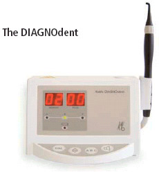
The first technology introduced for caries detection is the DIAGNOdent™ (KaVo, Biberach, Germany; KaVo America Corp, Lake Zurich, Ill). It uses infrared laser fluorescence and has been on the market outside of the United States since February 1998. It received US Food and Drug Administration approval in February 2000.
This instrument was developed for the detection and quantification of hypomineralization dental caries of occlusal and smooth surfaces. It uses a diode laser light source with a wavelength of 655 nm and I mW peak power.2 A fiber optic cable transmits the light to a hand-held probe with a fiber optic eye in the tip. The light is absorbed and induces infrared fluorescence by organic and inorganic materials. The emitted fluorescence is collected at the probe tip, transmitted through ascending fibers, and processed and presented on a display window as an integer in the range of 0 to 99. Increased fluorescence reflects carious tooth substance particularly with higher numerical values greater than about 20. The identification of the material responsible for the fluorescence is still under investigation but appears to be bacterial metabolites, particularly the porphyrins.3
A number of in vitro studies4-8 and a few in vivo studies9-12 have been conducted on the performance of the DIAGNOdent in the past 5 years. The results of the various in vitro studies indicate that the DIAGNOdent instrument is capable of detecting relatively advanced carious lesions with a very strong correlation between DIAGNOdent readings and histologic evidence of caries, but not the depth of the lesions into dentin. These studies also show that the instrument exhibits excellent reproducibility and good to excellent sensitivity. However, the results of the in vitro studies also indicate that the readings are influenced by several variables, including the degree of dehydration of the lesion, the presence of dental plaque, and the presence of various types of stain in occlusal fissures.
 The results observed in clinical investigations show significant differences in readings between different DIAGNOdent instruments with regard to occlusal caries resulting in questions on the selected value or reading of 20 or 25 to indicate the presence of caries. With a given instrument, the level of intra-operator agreement was generally very good while the inter-operator agreement typically ranged from good to very good.11 Also, DIAGNOdent readings increased linearly with clinical histologic measurements and, therefore, the instrument was reported12 to distinguish with good sensitivity between sound tooth structure or shallow enamel caries and deeper carious lesions in enamel or lesions extending into dentin. However, recent studies11-12 have not confirmed the close range of values related to the lesion extent reported previously.9 Eakle et al12 stated that the instrument is very good at indicating the presence of deeper lesions in enamel or into dentin that may not be apparent yet on radiographs but is unable to reliably indicate the depth of a dentinal lesion.
The DIAGNOdent instrument is particularly useful for confirming the presence of occlusal caries. Especially with the judicious use of the dental explorer to remove oral debris from occlusal pits and fissures, the DIAGNOdent is capable of confirming the presence or absence of caries in otherwise suspicious areas. Instrument readings greater than 20 or 25 reflect the likely presence of caries and greater readings generally reflect more extensive lesion progression, although, there does not appear to be a linear relation between the readings and the extent of the lesion. Prudent use of the instrument appears to be to identify early lesions that should be considered for preventive rather than restorative treatments.
DIFOTI™
Conventional clinical caries examinations routinely use transillumination to identify lesions located on the interproximal surfaces of the anterior teeth. Fiber optic transillumination (FOTI) has been available for clinical use for at least 30 years. It is designed to provide an intense light beam transmitted through a fiber optic cable to a specially designed probe to use transillumination on the proximal surfaces of posterior teeth. Repeated improvements have been made so it can be used on occlusal as well as proximal tooth surfaces. FOTI is commonly used, often in place of radiographs, in private practices in Europe. Numerous studies have been conducted comparing the use of FOTI to visual and radiographic examinations13 and a recent investigation indicates that dental practitioners are able to detect more proximal lesions with FOTI than with a visual examination, with or without the use of radiographs.14
DIFOTI is a further advancement of this technology where the visually observed images are captured using a digital charged couple device (CCD) camera and sent to a computer for analysis with dedicated algorithms. This instrument is commercially available relatively recently (Electro-Optical Sciences Inc, Irvington, NY) and very few investigations of its capabilities and limitations have been done. The results of an in vitro study15 that involved imaging of extracted teeth indicate that the instrument exhibited superior sensitivity for the detection of caries on all tooth surfaces as compared to radiological imaging. However, a recent in vitro investigation16 involving the development of artificial lesions over 14 weeks with intermittent imaging and radiographs at 2 week intervals indicates that the DIFOTI system was not able to determine the depth of lesions in contrast to radiographs. Although at least one clinical investigation of this instrument is in progress at Indiana University, there are no published reports of the clinical use of the instrument demonstrating its utility in practice. Nevertheless, on the basis of the prior reports using FOTI, it is reasonable to expect that DIFOTI will be at least as good as radiographs for detecting caries on interproximal tooth surfaces, although it may not be able to assess the depth of the lesion.
QLF
Without question, the most extensively investigated instrument available for the early detection of dental caries is QLF, (Inspektor Research Systems BV, Amsterdam, Netherlands). This instrument will be introduced in December 2003 by OMNII Oral Pharmaceuticals, West Palm Beach, Fla. This methodology began with the observation in 1978 by a Swedish dental scientist, Folke Sundström, that the use of a laser light of selected wavelength markedly enhanced the visibility of early noncavitated lesions.17 Subsequent investigations by this group confirmed the value of this approach for the early detection of caries clinically.18 Further studies established a relationship between the loss of fluorescence intensity with increasing mineral loss from the lesions compared to sound enamel.19,20 Numerous additional in vitro and in situ studies confirmed this important relationship between the amount of observed fluorescence and the mineral content of the lesions, thereby allowing the development of a system for truly assessing changes in either the progression or regression of carious lesions.21-25
 To facilitate clinical investigations as well as use in clinical practice, a small portable system was developed where the laser source is replaced by a regular light source and filter system.22 The illumination system consists of a 50 W Xenon microdischarge arc lamp provided with an optical band pass filter with a peak intensity of 370 nm (full width half measure of 80 nm) in order to produce blue light. The light illuminating the tooth is transported through a liquid-filled light guide. The fluorescent filtered images (high pass filter, l > 520 nm) are captured using a color CCD camera and a frame-grabber. Data are collected, stored, and analyzed by custom-made software. Since the first attempts at in vivo quantification of mineral changes in the enamel,21 the QLF method has undergone further development in both software and hardware.
During the past 5 years, a number of clinical investigations have been conducted and convincingly demonstrate the ability of the QLF instrument to detect early carious lesions and to accurately monitor either lesion progression or regression. The first of these studies26 monitored the changes in white spot lesions in orthodontic patients following the removal of the appliances and the institution of improved oral hygiene and a fluoride dentifrice. The remineralization of the lesions was impressively demonstrated within a few weeks. Another study27 in school-age children demonstrated that QLF detected five to 10 times more early lesions than conventional detection methods, was particularly useful for occlusal pits and fissures, and was reproducible.
A clinical study28 of white spot lesions in caries-active children in Sweden demonstrated significant remineralization of the lesions within 6 months following treatments with a fluoride varnish. An additional clinical study by this group29 involving multiple examiners demonstrated the repeatability and reproducibility of clinical QLF measurements. Quite recently, a team of Japanese scientists conducted a clinical trial using QLF to monitor changes in white spot lesions of children using a fluoride dentifrice and reported a significant decrease in the size of the lesions within 3 months of the 1 year test period.30 From these clinical studies, it is apparent that quantitative light fluorescence enhances the early detection of dental caries and is useful for monitoring the progression or regression of lesions. The only significant limitation is its inability to detect or monitor interproximal lesions.
Conclusion
Caries detection is entering a new era with new technologies capable of detecting lesions at an earlier stage of development and quantifying the impact of noninvasive professional fluoride treatments such as fluoride varnishes. DIAGNOdent appears to be most useful for the confirmation of the presence of caries in suspicious occlusal pits and fissures and the detection of deep dentinal lesions of the occlusal surface (so-called hidden caries). DIFOTI, pending further study, may have its greatest utility as a potential replacement for the use of bitewing radiographs for the detection of caries on interproximal surfaces. QLF is the most useful for the earlier detection of dental caries on occlusal, buccal, and lingual surfaces and the quantification of changes in early lesions associated with various preventive-oriented treatments. Caution should be exercised in examining newly erupted teeth with all these instruments, since these teeth are not yet fully calcified.
These new technologies are exciting for dental hygienists, not only as caries detection instruments but also to assess the impact of office treatments and to reinforce patient education.
References
- Pitts N. Advances in radiographic detection methods and caries management rationale. In: Stookey GK, ed. Early Detection of Dental Caries. Indianapolis: Indiana University School of Dentistry; 1996:39-50.
- Hibst R, Gall R. Development of a diode laser-based fluorescence caries detector. Caries Res. 1998;32:294.
- Hibst R, Paulus R. Molecular basis of red excited caries fluorescence. Caries Res. 2000;4:323.
- Ouellet A, Hondrum SO, Pietz DM. Detection of occlusal carious lesions. Gen Dent. 2002;50:346-350.
- Costa AM, Yamaguti PM, De Paula LM, Bezerra, AC. In vitro study of laser diode 655 nm diagnosis of occlusal caries. J Dent Child. 2002;69:249-253.
- Bamzahim M, Shi XQ, Angmar-Mansson B. Occlusal caries detection and quantification by DIAGNOdent and Electric Caries Monitor: in vitro comparison. Acta Odontol Scand. 2002;60:360-364.
- Alwas-Donowska HM, Plasschaert AJ, Suliborski S, Verdonschot EH. Reliability and validity issues of laser fluorescence measurements in occlusal caries diagnosis. J Dent. 2002;30:129-134.
- Lussi A, Francescut P. Performance of conventional and new methods for the detection of occlusal caries in deciduous teeth. Caries Res. 2003, 37:2-7.
- Lussi A, Megert B, Longbottom C, Reich E, Francescut P. Clinical performance of a laser fluorescence device for detection of occlusal caries lesions. Eur J Oral Sci. 2001; 109:14-19.
- Anttonen V, Seppa L, Hausen H. Clinical study of the use of the laser fluorescence device DIAGNOdent for detection of occlusal caries in children. Caries Res. 2003;37:17-23.
- Tranæus S, Lindgren LE, Karlsson L, Angmar-Mansson, B. Evaluation of the in vivo performance of the DIAGNOdent device. In: Stookey GK, ed. Early Detection of Dental Caries. 3rd ed. Indianapolis: Indiana University School of Dentistry. In press.
- Eakle WS, Gansky SA, Zhan L, Featherstone JDB. Clinical evaluation of the DIAGNOdent device. In: Stookey GK, ed. Early Detection of Dental Caries. 3rd ed. Indianapolis: Indiana University School of Dentistry. In press.
- Pine CM. Fiber-optic transillumination (FOTI) in caries diagnosis. In: Stookey GK, ed. Early Detection of Dental Caries. Indianapolis: Indiana University School of Dentistry; 1996:51-65.
- Davies GM, Worthington HV, Clarkson JE, Thomas P, Davies RM. The use of fiber-optic transillumination in general dental practice. Br Dent J. 2001;191:145-147.
- Schneiderman A, Elbaum M, Shultz T, Keem S, Greenebaum M, Driller J. Assessment of dental caries with Digital Imaging Fiber-Optic TransIllumination (DIFOTI™): in vitro study. Caries Res. 1997;31:103-110.
- Young DA, Featherstone JDB. Comparing digital imaging fiber-optic trans-illumination, F-speed radiographic film, and polarized light microscopy. In: Stookey GK, ed. Early Detection of Dental Caries. 3rd ed. Indianapolis: Indiana University School of Dentistry. In press.
- Bjelkhagen H, Sundstrom F. A clinically applicable laser luminescence method for the early detection of dental caries. IEEE J Quantum Electronics. 1981;80:120-122.
- Sundstrom F, Fredriksson K, Montan S, Hafstrom-Bjorkman U, Strom J. Laser-induced fluorescence from sound and carious tooth substance: spectroscopic studies. Swed Dent J. 1985;9:71-80.
- Hafstrom-Bjorkman U, Sundstrom F, de Josselin de Jong E, Oliveby A, Angmar-Mansson B. Comparison of laser fluorescence and longitudinal microradiography for quantitative assessment of in vitro enamel caries. Caries Res. 1992;26: 241-247.
- Angmar-Mansson B, ten Bosch JJ. Optical methods for the detection and quantification of caries. Adv Dent Res. 1987; 1:14-20.
- de Josselin de Jong E, Sundstrom F, Westerling H, Tranæus S, ten Bosch JJ, Angmar-Mansson B. A new method for in vivo quantification of changes in initial enamel caries with laser fluorescence. Caries Res. 1995;29:2-7.
- al-Khateeb S, ten Cate JM, Angmar-Månsson B, et al. Quantification of formation and remineralization of artificial enamel lesions with a new portable fluorescence device. Adv Dent Res. 1997;11:502-506.
- al-Khateeb S, Oliveby A, de Josselin de Jong E, Angmar-Mansson B. Laser fluorescence quantification of remineralization in situ of incipient enamel lesions: influence of fluoride supplements. Caries Res. 1997;31:132-140.
- Ferreira Zandona AG, Analoui M, Schemehorn BR, Eckert GJ, Stookey GK. Laser fluorescence detection of demineralization in artificial occlusal fissures. Caries Res. 1998;32:31-40.
- Lagerweij M, van der Veen M, Ando M, Lukantsova L, Stookey GK. The validity and repeatability of three light-induced fluorescence systems: an in vitro study. Caries Res. 1999;33:220-226.
- al-Khateeb S, Forsberg CM, de Josselin de Jong E, Angmar-Mansson B. A longitudinal laser fluorescence study of white spot lesions in orthodontic patients. Am J Orthod Dentofacial Orthop. 1998;113:595-602.
- Ferreira Zandona AG, Isaacs RL, van der Veen M, Stookey, GK. Indiana pilot clinical study of quantitative light fluorescence. In: Stookey GK, ed. Early Detection of Dental Caries. 2nd ed. Indianapolis: Indiana University School of Dentistry; 2000:219-230.
- Tranæus S, al-Khateeb S, Bjorkman S, Twetman S, Angmar-Mansson B. Application of quantitative light-induced fluorescence to monitor incipient lesions in caries-active children. A comparative study of remineralisation by fluoride varnish and professional cleaning. Eur J Oral Sci. 2001;109:71-75.
- Tranæus S, Shi XQ, Lindgren LE, Trollsas K, Angmar-Mansson B. In vivo repeatability and reproducibility of the quantitative light-induced fluorescence method. Caries Res. 2002;36:3-9.
- Kambara M, Uemura M, Doi T. Results of clinical trial of fluoride dentifrices using QLF. In: Stookey GK, ed. Early Detection of Dental Caries. 3rd ed. Indianapolis: Indiana University School of Dentistry. In press.
From Dimensions of Dental Hygiene. October 2003;1(6):12-13, 15.
