Technique Focus
Due to their superior accessibility and adaptability, mini-bladed Gracey curets have opened a new chapter in the history of root debridement.
In recent years, modifications and advancements have abounded in hand instrument design. To chronicle these advancements, this will be the first in a series on the design and use of some of the most innovative and effective developments in hand instruments.
Mini-bladed Gracey curets are modifications of the extended shank Gracey curets. Their terminal shanks are 3 mm longer and their blades are thinner and half the length of standard and extended shank Gracey curets. (Figure 1). The shorter, thinner blade allows easier insertion and adaptation in deep, narrow pockets; furcations; developmental depressions; line angles; and deep, tight pockets on facial, lingual, or palatal surfaces. In areas where root morphology or tight tissue prevent full insertion of the standard or extended shank Gracey blade, the mini-bladed Gracey curets can be used with vertical strokes, with minimal tissue distention, and without tissue trauma.
Mini-bladed Gracey curets have substantially improved the degree to which deep subgingival deposits of calculus and biofilm can be removed. This is due to superior access and adaptation of their blades to anatomical contours exposed by moderate to severe clinical attachment loss. In the past, the only solution in difficult-to-access areas was to use standard Gracey curets with a short, toe-down, horizontal stroke.

Micro mini-bladed Gracey curets are the latest modification of mini-bladed Gracey curets. They are now being used in Germany and are not currently available in the United States (Figure 2). These instruments are half the size of the currently available mini-bladed Gracey curets. They allow better access to furcations and very tight, narrow pockets and are ideal for subgingival use with the dental endoscope. This trend toward miniaturization of instruments facilitates non-surgical treatment of areas that were previously difficult or impossible to reach with standard hand instruments.

Rigidity

Mini-bladed Gracey curets are available in both finishing and rigid shanks. If a mini-bladed Gracey curet does not feel sturdy enough during scaling, switch to one with a more rigid shank. These stronger, thicker shanked mini-bladed Gracey curets provide the necessary rigidity to prevent flexing of the shank while scaling tenacious calculus. Although many
manufacturers offer rigid mini-bladed Gracey curets, some process their metals specially to provide varying degrees of hardness, sharpness, flexibility, or rigidity. The EverEdge™ technology from Hu-Friedy, Chicago, offers a very hard steel that requires less sharpening. The XP™ instruments from American Eagle, Missoula, Mont, require no sharpening due to its special process of impregnating titanium nitrate into stainless steel by surface engineering. Replacement is required when the instrument becomes dull after extended use. Paradise Dental Technologies, Missoula, Mont, and G. Hartzell Co, Concord, Calif, also produce rigid mini-bladed Gracey curets with hard, sharp blades that are helpful for scaling tenacious or burnished calculus.

Technique Differences
The technique used is generally the same as the Gracey curets except for the following differences:
- Mini-bladed Gracey curets should not be used routinely for all tooth surfaces in place of standard Gracey or extended shank Gracey curets. Rather, they should be used instead of conventional Gracey curets in areas of difficult access such as furcations; line angles; and deep, tight, narrow pockets, especially on facial and palatal surfaces (Figure 1).
-

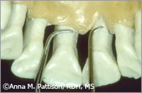
Figure 6a and b. Mini-bladed Gracey 5/6 (a) and 13/14 (b) adapted to the buccal furcation of a maxillary molar. Large diameter ergonomic handles are recommended for any mini-bladed instrument because the larger diameter allows better control of the small blades.
- Mini-bladed Gracey curets can be positioned with the toe directed either mesially or distally for vertical strokes on buccal or lingual surfaces. On posterior teeth, the blade adapts better to these surfaces when the toe is directed distally. Vertical strokes are then activated from the mesial line angle toward the distal line angle (Figure 3).
- Use rigid mini-bladed Gracey curets for calculus removal. Use the thinner shanked, finishing mini-bladed Gracey curets for root debridement of biofilm and/or light scaling of periodontal maintenance patients with tight pockets. It is best to select the appropriate set based on the specific needs of the patient.
- When using mini-bladed Gracey curets for calculus removal, use intraoral finger rests close to the working area. For deep pockets on the maxillary arch, try moving in the range of 1 o’clock to 5 o’clock on the left side of the chair. Recline the back of the chair so that the patient’s head is as far back as possible and then ask the patient to lift his or her chin and open wide. From this position, it is possible to use intraoral fulcrums to scale deep pockets on the maxillary posterior teeth with the same techniques that you routinely use for the mandibular teeth (Figure 4).
- When performing light scaling or deplaqueing, either intraoral or extraoral rests may be used. Extraoral rests are usually necessary to gain access to deep pockets in areas such as the maxillary second and third molars (Figure 5).
- Mini-bladed Gracey curets are generally used with straight vertical strokes. They may also be used with oblique or horizontal strokes but due to the shortness of the blade, these strokes might not extend far enough subgingivally unless the tissue is very retractable. Horizontal strokes with mini-bladed Gracey curets are most effective when limited to use along the cementoenamel junction (CEJ) area or in developmental depressions just below the CEJ.
- For instrumentation of maxillary furcations, the mini-bladed Gracey 13/14 curet is the most versatile. A front position (7 o’clock to 9 o’clock) allows access to buccal, distal, and mesial furcations of all of the maxillary molars with either intraoral or extraoral fulcrums. For buccal furcations, use a mini-bladed Gracey 11/12 or 5/6 for the mesial surface of the disto-buccal root and a mini-bladed Gracey 13/14 for the distal surface of the mesio-buccal root (Figure 6). For maxillary distal or mesial furcations, it is possible to scale both sides of these furcations with the mini Gracey 13/14 by using opposite arch or extraoral fulcrums.
- 9. For instrumentation of mandibular furcations, a front position requires that you use the combination of the mini-bladed Gracey 11/12 and 13/14 with intraoral finger rests to scale any buccal or lingual furcation.
By positioning the patient more upright and moving to a standing back position, an opposite arch or maxillary extraoral fulcrum can be used. With this approach, any mini-bladed Gracey curet can scale both the mesial and distal aspects of mandibular furcations.
Mini-bladed Gracey curets provide unprecedented access to difficult areas for non-surgical root debridement. In areas such as line angles, furcations, and narrow, curved, facial, or palatal root surfaces, the miniature blades of these curets provide superior adaptation with excellent tactile sensitivity. For this reason, they are ideal for final scaling and root planing following either ultrasonic or traditional hand scaling.
This article was adapted from Pattison A, Matsuda S, Pattison G. Periodontal Instrumentation. 3rd ed. Upper Saddle River, NJ: Prentice Hall. In press.
From Dimensions of Dental Hygiene. February 2006;4(2):28-30.
