 ROBERTPRZYBYSZ/ISTOCK/THINKSTOCK
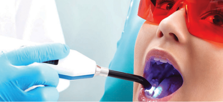
Sealant Success
Dental sealants remain an integral part of the caries prevention armamentarium.
Dental sealants were introduced in the 1960s to prevent dental caries.1 In the 1970s and 1980s, caries rates declined in the United States, most likely due to the widespread use of topical and systemic fluoride.1 During this time, however, sealants were not widely used and occlusal caries rates remained high.1
Today, occlusal caries rates are higher than smooth surface caries rates among children and adolescents.2 While sealant usage has grown, many high-risk children do not have access to this valuable caries prevention tool.
Along with changing caries rates and varying levels of sealant usage, the materials that comprise dental sealants have significantly evolved. Bonding agents were introduced in the early 1990s, and self-etching agents were launched in the 2000s.3 Self-etching sealants eliminated the separate etchant step initially required in the sealant placement process. However, low retention rates prevented the American Dental Association from advocating their use.
Modern pit and fissure sealants are typically resin-based cements or glass-ionomer cements.1 Most resin sealants are hydrophobic and are either urethane dimethacrylate or bisphenol A-glycidyl methacrylate monomers.4 Resin sealants typically have a wide range of characteristics, including being tinted or clear in color, filled (with glass or quartz particles for wear resistance) or unfilled, chemically activated or light-activated to polymerize, and fluoride releasing. Resin-based materials that can be placed in a moist field are also available. This supports a quick application process, which can be particularly helpful when placing sealants in young children.5,6 Resin-based sealants have longer retention rates than glass ionomer sealants.2
Glass ionomer sealant materials contain an acidic liquid that reacts with a glass powder. They are hydrophilic and suitable for placement in a moist environment. They are also fluoride releasing. Due to decreased retention rates, however, glass ionomer sealants are most often used as interim sealants.2
TECHNIQUE TIPS FOR OPTIMUM RETENTION
Retention of dental sealants is key to their effectiveness in caries prevention and is often impacted by clinician skill and technique. Each sealant material manufacturer has specific guidelines for sealant placement, and following these guidelines is critical for the success of sealant placement.
Gray et al7 reviewed the existing literature on tooth preparation prior to etchant and sealant placement. They found that toothbrushing or mechanical cleaning with a handpiece is acceptable prior to acid etching. When cleaning the tooth surface prior to acid etching, however, an oil-based agent may impede the action of the acid-etch, creating a microscopically roughened enamel surface that hampers sealant retention.8 A nonoil-based agent—such as fine flour of pumice mixed with water—should be considered.4 Rinsing the tooth of the cleansing agent and debris is necessary. An explorer tip can be used with very light pressure to mechanically debride the fissure of the residual agent. However, using an explorer with too much pressure can cavitate a surface, so caution is recommended.
Moisture contamination accounts for most traditional resin-based sealant failures.9 For sealants requiring a dry field, prior to conditioning the tooth surface with acid etchant, the field should be isolated to prevent moisture contamination. The use of isolation techniques, such as cellulose triangles and cotton roll holders, is essential to maintaining a dry field. In addition, the use of a four-handed placement technique and high-volume evacuation to mitigate saliva contamination may enhance sealant placement.5,9
Proper etching technique is critical to effective sealant placement. Clinicians need to be aware of the proper etching time for the product they’re using. If a liquid etchant is used, the etchant must be continuously dabbed, not rubbed onto the tooth surface. This will maintain the pattern of the enamel rods without causing damage.4 Properly rinsing any residual etchant from the tooth is necessary for the retention of the sealant in the fissural anatomy. Once rinsed, the etched area of the tooth should appear frosted to indicate successful conditioning of the tooth surface.4 During resin sealant placement, the material should not be over-manipulated, as this can create air bubbles in the sealant surface. Once the sealant has been applied, visible light polymerization can take place.
SEALANT EFFECTIVENESS AND ADVOCACY
The occlusal surface is eight times more likely to decay than smooth enamel surfaces.10 This fact coupled with the anatomical features of occlusal surfaces make them ideal for dental sealants. According to Hegde and Coutinho,11 completely erupted, caries-free permanent teeth are best suited for sealant placement. However, clinical practice guidelines released by the American Dental Association Council on Scientific Affairs in 2008 and updated in 2016 recognize the secondary prevention benefits afforded by dental sealants, and recommend sealant placement on early noncavitated caries lesions.2,5 Research shows that dental sealants are not only effective in preventing decay, but also in arresting the caries process in its early stages.2 By blocking the food source, the bacteria in biofilm are robbed of the nutrients they need to begin the demineralization process. The clinical practice guidelines also identify the need to seal both primary and permanent molars to prevent the caries process.5
Benefits of dental sealants include cost savings, time savings, and tooth preservation. By preventing or arresting decay, potentially expensive or extensive restorative procedures can be avoided and less time is spent in the dental chair for both patients and clinicians. The treatment of decay requires excavation of the caries lesion, which involves removal of tooth structure prior to restoration placement. By preserving the enamel during sealant placement, the tooth structure is kept intact.
CONCLUSION
While dental sealants are a proven strategy in the caries prevention armamentarium, their usage leaves much to be desired. According to a recent US Centers for Disease Control and Prevention report, approximately 60% of children age 6 to 11 don’t have dental sealants.12 The report also noted that school-age children without sealants experience almost three times more caries lesions than children with sealants.12 Oral health professionals can improve these dismal usage rates by educating their patients about sealant benefits, remaining up to date on the latest developments in the sealant-placement technique, and supporting school-based sealant programs. By significantly reducing caries rates, oral health professionals can help improve their patients’ oral and overall health.
REFERENCES
- Ahovuo-Saloranta A, Forss H, Walsh T, et al. Sealants for preventing dental decay in the permanent teeth. Cochrane Database Syst Rev. 2013;3:CD001830.
- Wright J, Crall J, Fontana M, et al. Evidence-based clinical practice guidelines for the use of pit-and-fissure sealants. J Am Dent Assoc. 2016;147:672–682.
- Tandon V, Lingesha R, Yadav V. Effect of adhesive application on sealant success: a clinical study of fifth and seventh generation adhesive systems. J Dent (Tehran). 2015;12:712–719.
- Powers J, Wataha J. Dental Materials: Properties and Manipulation. 10th ed. St. Louis: Mosby; 2013.
- Beauchamp J, Caufield P, Crall J, et al. Evidence-based clinical recommendations for the use of pit and fissure sealants: a report of the American Dental Association Council on Scientific Affairs. J Am Dent Assoc. 2008;139:257–268.
- Hagel N, Vannah D. Seal away caries risk. Dimensions of Dental Hygiene. 2015;13(6):34–36.
- Gray S, Griffin S, Malvitz D, Gooch B. A comparison of the effects of toothbrushing and handpiece prophylaxis on retention of sealants. J Am Dent Assoc. 2009;140:38–46.
- Wilkins E. Clinical Practice of the Dental Hygienist. 12th ed. Philadelphia: Lippincott, Williams, and Wilkins; 2016.
- Griffin S, Jones K, Gray S, Malvitz D, Gooch B. Exploring four-handed delivery and retention of resin-based sealants. J Am Dent Assoc. 2008;139:281–289.
- Kumaran P. Clinical evaluation of the retention of different pit and fissure sealants: a 1-year study. Int J Clin Pediatr Dent. 2013;6:183–187.
- Hegde R, Coutinho R. Comparison of different methods of cleaning and preparing occlusal fissure surface before placement of pit and fissure sealants: an in vivo study. J Indian Soc Pedod Prev Dent. 2016;34:111–114.
- Centers for Disease Control and Prevention. Dental sealants prevent cavities. Vital Signs. Available at: cdc.gov/vitalsigns/dental-sealants/index.html. Accessed October 19, 2016.
From Dimensions of Dental Hygiene. November 2016;14(11):24,26.
