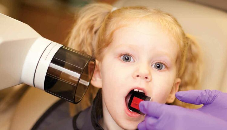
Eliminate Radiographic Retake Exposures for Patients With Special Needs
Altering techniques to obtain diagnostic quality images in all patients—including children and those presenting with challenges—is key to the safe and effective capture of radiographs.
“As low as reasonably achievable,” or ALARA, is a critical radiation safety principle, as ionizing radiation can cause biological health risks. Organizations, such as the American Dental Association, American Dental Hygienists’ Association, and American Academy of Pediatric Dentistry, promote ALARA.1–3 Evidence-based selection criteria guidelines developed by an expert panel of professionals should be used when making decisions about the type, frequency, and quantity of dental radiographs to be taken for adults, adolescents, and children.4
Dental radiographic images are essential to diagnosing oral disease. As such, dental radiographers must be able to capture quality images while eliminating the need for retake, which supports ALARA. While many factors promote ALARA, including digital vs film-based imaging, digital image receptor type, collimation, exposure settings, and thyroid shielding, exposing quality radiographic images on the first attempt is key.5 However, eliminating retake exposures can be challenging as many patients face special needs during radiographic examinations. For example, pediatric patients typically have low palatal vaults or shallow palates due to their smaller size and growth and development.6 In addition, many children and adults have a hypersensitive gag relax. Patients with conditions such as autism spectrum disorder (ASD); patients who use a wheelchair; and those who experience uncontrolled movements caused by disorders, such as Parkinson disease and Alzheimer disease, also present challenges to radiographic imaging techniques.
HYPERSENSITIVE GAG REFLEX
While everyone has a gag reflex, the presence of a hypersensitive gag reflex may make placing and obtaining diagnostic images difficult.6 Approximately 6% of children and adolescents and 49% of adults experience a hypersensitive gag reflex associated with dental treatment.7,8 Often, the soft palate or posterior third of the tongue is stimulated when placing and exposing intraoral radiographs, triggering the gag reflex. Techniques to reduce hypersensitive gag reflex often focus on decreasing psychogenic and tactile stimuli. To mitigate psychogenic stimuli, distraction techniques, such as lifting the leg and concentrating on keeping it lifted, can be implemented. Empathize with the patient and avoid bringing up gagging or “suggesting” comfort unless the patient discusses these items first, as this may exacerbate the problem. Breathing techniques, such as breathing through the nose, humming, or holding the breath for the duration of the exposure, may alleviate the gag reflex, as well.6 To reduce tactile sensitivity, avoid sliding the image receptor across the tissues and mucosa, and consider the use of an image receptor cushion. Manually stimulating the palatal vault and tongue with a toothbrush is effective in desensitizing the tissues to make placement of an image receptor less foreign and less likely to trigger the gag.6,9 Additionally, confusing the senses by rinsing with cold water or antiseptic mouthrinse, or placing salt on the tip or middle of the tongue can help make intraoral image receptor placement more tolerable.6
In extreme cases, alternative methods to manage the hypersensitive gag reflex may be necessary. Extraoral techniques, where the sensor is stabilized outside of the patient’s mouth, may assist in obtaining reasonably diagnostic images when intraoral radiographic images are impossible to obtain.10 Additionally, acupuncture on specific pressure points may help to decrease the gag response, as well as topical anesthetic and advanced sedation.11–16 These alternative techniques, however, should be used as a last resort as acupuncture may not be suitable for all patients.12 The use of sedation also poses risks and may increase patient anxiety. Finally, the use of an extraoral imaging method could result in image magnification and distortion, reducing diagnostic ability.10
TORI OR SHALLOW PALATES
The presence of a tori or shallow palate hinders the radiographer’s ability to place an intraoral image receptor parallel to the teeth of interest. While the paralleling technique is the most optimal to reduce retake errors during intraoral imaging, the bisecting technique is acceptable in cases where parallel placement is not possible. For the bisecting technique, radiographers place the image receptor at an angle very close to the teeth of interest, and alter vertical angulation to be perpendicular to an imaginary line formed by the plane of the image receptor and plane of the teeth of interest.6 With the bisecting technique, some image distortion can result; however, minimal distortion is considered acceptable for diagnosing oral disease with this technique.6 Research comparing the paralleling technique and bisecting technique on 380 images of simulated fragmented skull remains using a handheld device found significantly more vertical angulation errors on the bisecting technique, requiring a retake exposure. Radiographer skill in the bisecting technique for situations of tori or shallow palate is important to reduce retake exposures. Allowing time for radiographers to practice the bisecting technique through simulation is recommended.17
Vertical angulation may need to be slightly increased to accommodate image receptors in children with a low palatal vault. A smaller image receptor size and modifications to image receptor positioners may be necessary for the pediatric patient. For large mandibular torus, the radiographer should attempt to position the image receptor between the torus and tongue. An image receptor cushion may make placement near the torus more comfortable as the image receptor may feel uncomfortable on the protruding bone.6
SPECIAL NEEDS DISORDERS OR CONDITIONS
Challenges can arise for patients who have difficulty remaining still for radiographic imaging as movement causes distorted, undiagnostic images, resulting in the need for retake exposures. With handheld imaging, the radiographer remains with the patient during all exposures. While handheld radiography equipment is ideal for use in situations where wall-mounted dental X-ray devices are not accessible—such as mobile dental clinics and outreach events where dental hygiene practitioners may provide oral hygiene assessment and treatment—the radiographer’s ability to hold the handheld equipment during exposure could provide increased stability for patients with special needs. Handheld radiographic imaging for intraoral radiographic images may help radiographers achieve optimal results when involuntary movement occurs due to special needs. Additionally, a handheld unit may be useful in treating patients who use wheelchairs and cannot transfer to the dental chair, or the equipment cannot access the patient while in the wheelchair. Using a handheld device can also assist with special needs patients including those with ASD or other sensory processing disorders, so that the radiographer can provide clear, calm instructions throughout the procedure and reduce the amount of time needed to obtain a final image. Handhelds eliminate the need for the radiographer to leave the room to make an exposure, as images are exposed directly from the handheld device after the image receptor is placed in the correct position.18
Radiographers using handheld radiographic equipment should be knowledgeable on the lowest exposure settings to produce acceptable images and apply safe practices for the operator and patient. While handhelds are equipped with a backscatter ring shield around the position indicating device and inherent shielding, operators should don an operator lead apron with thyroid shielding when the device is in any position away from operator mid torso height or vertical angulation at 0° with the device parallel to the floor. The increased vertical angulations for periapical images require patients to move their head upward for mandibular periapical images and downward for maxillary periapical images for optimal operator protection; however, this could be challenging for patients with special needs.18,19 To assist in determining accurate angulations for high-quality images with handhelds and the paralleling technique, manufacturers of image receptor holding devices have shortened the metal positioning arm for use with handheld radiographic equipment. Altered equipment should be used together with handheld devices to assist with required angulations.18
Patients with ASD and other special needs that affect sensory processing often benefit from practice at home before the radiographic procedure, as ASD can impact communication and social interaction skills.20 This may include a “practice kit” for at-home use, such as a disposable bite block so the patient can rehearse holding the receptor while a caregiver counts or distracts the patient.21 Watching videos or reading handouts that model a radiographic procedure may make the procedure seem more familiar and increase compliance and cooperation. Additionally, the weight of two radiographic aprons, sunglasses, ear plugs, and a calm and quiet environment may assist in obtaining radiographs.21
CONCLUSION
Obtaining diagnostic quality images on patients with special needs may present challenges. Basic radiographic procedures and techniques may need to be altered to reduce retakes and adhere to ALARA principles. Patients with special needs may be anxious about radiographic treatment, such as those with a hypersensitive gag reflex and ASD. Therefore, establishing a rapport and displaying empathy increases cooperation and the likelihood of obtaining the highest quality images. Radiographers using handheld imaging devices to assist with the radiographic procedure must ensure all safety mechanisms are implemented for operator safety.
Oral health professionals should seek additional education and knowledge in radiographic techniques to safely and effectively treat patients with special needs to support the ultimate goal of achieving diagnostic quality radiographic images on the first attempt. Continuing education courses focusing on treating special needs populations can assist in this effort.
REFERENCES
- White SC, Scarfe WC, Schulze RK, et al. The Image Gently in Dentistry campaign: promotion of responsible use of maxillofacial radiology in dentistry for children. Oral Surg Oral Med Oral Path Oral Radiol Endod. 2014;118:257–261.
- Bruhn AM, Newcomb TL, Tolle SL. Ensuring safe practice in dental radiology. Dimensions of Dental Hygiene. 2015;13(12):30–33.
- Wenzel A, Moystad A. Work flow with digital intraoral radiography: a systematic review. Acta Odontol Scand. 2010;68:106–114.
- American Dental Association: Council on Scientific Affairs. Dental Radiographic Examinations: Recommendations for Patient Selection and Limiting Radiation Exposure. Available at: ada.org/~/media/ADA/Member%20Center/FIles/Dental_Radiographic_Examinations_2012.ashx. Accessed March 16, 2021.
- Bruhn A, Suedbeck J. The safe use of radiography in children. Dimensions of Dental Hygiene. 2017;15(2):24–27.
- Thomson E, Johnson O. Essentials of Dental Radiography for Dental Assistants and Hygienists. 10th ed. Upper Saddle River, New Jersey: Pearson Education Inc; 2018:436.
- Randall CL, Shulman MS, Grant P, Crout BS, Richard J, McNeil DW. Gagging and its associations with dental care–related fear, fear of pain and beliefs about treatment. J Am Dent Assoc. 2014;145:452–458.
- Katsoudas M, Provatenou E, Arapostathis K, Coolidge T, Kotsanos N. The Greek version of the Gagging Assessment Scale in children and adolescents: psychometric properties, prevalence of gagging, and the association between gagging and dental fear. Int J Paediatr Dent. 2017;27:145–151.
- Neumann JK, McCarty GA. Behavioral approaches to reduce hypersensitive gag response. J Prosthet Dent. 2001;85:305.
- Silva MHC, Coelho MS, Santos MFL, De Lima CO, Campos CN. The use of an alternative extraoral periapical technique for patients with severe gag reflex. Case Rep Dent. 2016;3206845:1–5.
- Okar S, Kaviani NR, Soltani P, Ahmadi A, Haghighat A. Evaluation of the effects of acupuncture on P6 and anti-gagging acupoints on the gag reflex. Dental Hypotheses. 2015;6(1):19–22.
- Vachiramon A, Wang WC, Vachiramon T. The use of acupuncture in implant dentistry. Implant Dent. 2004;13:58–64.
- Fiske J, Dickinson C. The role of acupuncture in controlling the gagging reflex using a review of ten cases. Br Dent J. 2001;190:611–613.
- Lu DP, Lu GP, Reed JF 3rd. Acupuncture/acupressure to treat gagging dental patients: a clinical study of anti-gagging effects. Gen Dent. 2000;48:446–552.
- De Veaux CKE, Montagnese TA, Heima M, Aminoshariae A, Andre M. The effect of various concentrations of nitrous oxide and oxygen on the hypersensitive gag reflex. Anesth Prog. 2016;63:181–184.
- Yamamoto T, Fujii-Abe K, Fukayama H, Kawahara H. The effect of adding midazolam to propofol intravenous sedation to suppress gag reflex during dental treatment. Anesth Prog. 2018;65:76–81.
- Bruhn A, Newcomb T, Giles B. Evaluating imaging techniques for intraoral forensic radiography with the dental hygienist as part of the forensic radiology yeam. J Forensic Identif. 2016;66:22–36.
- Bruhn A, Lintag K. Technique tips for handheld radiography. Perspectives on the Midlevel Practitioner. 2018;(Special Supplement):28–31.
- Aribex Inc. Portable X-ray System for Intraoral Radiographic Imaging User Manual. Available at: q9bgh9q08416907ck9fxol3z-wpengine.netdna-ssl.com/wp-content/uploads/NOMAD_Classic_MP-0013_Rev-G.pdf. Accessed March 16, 2021.
- Elmore J, Bruhn A, Bobzien J. Research: interventions for the reduction of dental anxiety and corresponding behavioral deficits in children with autism spectrum disorder (ASD). J Dent Hyg. 2016;90:111–120.
- Dailey JC, Brooks JK. Autism spectrum disorder: Techniques for dental radiographic examinations. J Dent Hyg. 2019;93:35–41.
From Dimensions of Dental Hygiene. April 2021;19(4):16,18,21.

