Digital Dental Imaging
Incorporating digital photography into the dental operatory.
Dental photography is an important adjunct to dental health care delivery. Intraoral photography with 35 mm slides and printed photos was time consuming, technique sensitive, and expensive. Photographs had to be exposed and then developed before the results could be evaluated—making the photography session a trial run where photos that turned out poorly had to be retaken. Many dental professionals did not feel the time and expense were worth the effort.
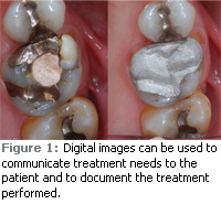 Digital photography is inexpensive and the results are available immediately, which make digital dental imaging an exciting treatment option. Digital photos can be used for documentation, patient education, and professional presentations. A simple digital occlusal photograph can illustrate the patient’s current condition on a computer monitor in the treatment room and can even be displayed on a monitor attached to the patient’s chair. The high quality of current digital images permits users to zoom in on single teeth to point out caries, failing margins, cracks, or other treatment needs. A picture can clearly communicate needs that many patients have a difficult time understanding through verbal explanations (Figure 1). These images also create valuable records of the patient’s current condition prior to the start of dental restorative procedures. Documentation is key to satisfying insurance claims and patient complaints concerning the care received during their treatment.
Confidentiality is an issue, and any photo in which the patient can be identified must have the appropriate legal release or undergo alteration. The digital file must also be stored in a protected, secure electronic environment. Fortunately, most photos of individual teeth or even quadrants of teeth do not have any identifying information that would lead to patient identification.
Photos of dental techniques and treatments can be used in booklets, educational pamphlets, or newsletters to explain simple techniques or illustrate esthetic cases to current or prospective patients. Slide shows can be arranged using photographic software to present a procedure from beginning to end. These slide shows can then be displayed on digital picture frames in the waiting room or in more sophisticated settings on larger displays such as LCD televisions.
ADVANTAGES
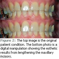 Intraoral digital photography is similar to film photography; the camera, lens, and flash equipment are basically the same as the older 35 mm slide photography systems. The distinct advantage of digital imaging is that the cost per image is virtually nothing after the initial investment, and the results can be used immediately. Photos can be downloaded to computers in the dental treatment room, treatment planning area, or the dentist’s office. Networked computers allow the movement of digital files to the appropriate venue. With some practice, digital images can be manipulated quickly to show proposed esthetic changes to patients without altering the physical tooth structures (Figure 2).
Photos can be printed for patients to take home while they consider the proposed treatment. These files can be easily stored in patient records if electronic patient record technology is in use. Most office management software has provisions for adding digital images to patient records. Even if electronic records are not used, careful planning of the storage filing system can lead to easy retrieval and use of the photographs. Many digital photo software packages include album storage and filing systems that allow users to specify how the files are named and stored. Most album software allows the use of keyword assignment to make searching for photos of a particular subject simple as the digital image library grows larger.
EQUIPMENT
To use digital imaging in the office, the first step is to choose the equipment that provides the most versatility and quality for the least outlay both financially and from a training and implementation aspect. There are two options: the single lens reflex (SLR) camera with a macro zoom lens and ring or ring/point combination flash and the intraoral wand camera.
The smaller “point and shoot” cameras have a number of limiting factors that make them difficult to use in dental imaging. The lens system of smaller digital cameras is not capable of using true optical properties to achieve the magnification necessary for intraoral photography. They use digital manipulation or auxiliary magnifying lenses to make the object appear larger but this manipulation reduces the quality of the image to an unacceptable level. Most of these cameras are also not capable of producing focused images over the depth of a typical dental photograph since they are designed for recreational photography. When using the SLR camera, special lensmounted flash units are required to produce consistent light, and these small cameras have no mounting system to provide for this feature.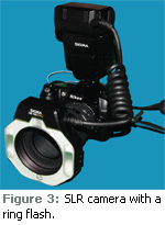
The SLR camera with the ring flash provides high quality digital images (Figure 3). A typical SLR camera is available for $500 to $600 with more expensive models available if more features are desired. These extra features are not typically used in dental photography so only consider the more expensive models if the camera will be used beyond the dental office. Macro-zoom lenses with a 105 mm length are most commonly used in dental photography and they cost approximately $400 to $600. Ring or ring/point flash systems usually range from $350 to $700.
Additional equipment is needed to take intraoral photographs with the SLR camera, including intraoral mirrors, cheek retractors, and photographic software to manage the digital images. Intraoral mirrors now have handles attached to make placement easier during the photo sessions. Occlusal and buccal mirrors of various sizes are recommended. Front surface glass mirrors produce the best images. These mirrors are more expensive but can be heat sterilized and are worth the investment. Retractors come in all types of styles and materials. There are self-retracting styles, clear plastic, stainless steel, and many other variations. Finding the retractor that works best is a personal discovery and clinicians may need to try a few different styles to make that determination.
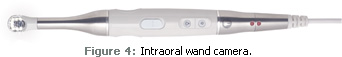
Intraoral wand cameras, which are used inside of the mouth and require barrier coverage, provide excellent visualization of the oral cavity (Figure 4). The main advantages of this type of intraoral camera are ease of use and the immediate viewing ability of images. They are well designed for use in difficult to reach posterior areas and where light is not easily provided. Intraoral wand cameras are equipped with USB connections or docking stations, providing portability. They offer their own high intensity light source as well as magnification ability up to 40x to 50x. Intraoral wand cameras are primarily designed to take one shot of one tooth at a time to maximize the effectiveness of their optical systems. The cost of intra – oral wand cameras is a factor with a high-end model costing between $3,500 and $5,500.
In order to incorporate digital images into the dental practice, imaging software is necessary. Its use requires a learning curve, like any new skill. Many packages come with their own tutorials to help the user get started and then grow more proficient at manipulation, storage, and use of the photo images. Many software companies offer online or in-person courses about the use of their software. As with most new tools, only practice and patience will lead to consistent results.
From Dimensions of Dental Hygiene. September 2009; 7(9): 24, 26.
