A Closer Look At Ultrasonic Scalers
Keeping up to date on the various ultrasonic units and tips available will help you make the most efficacious choice when using ultrasonic instrumentation with your patients.
On September 24, 2005, the American Academy of Periodontology (AAP) held its Third Annual Dental Hygiene Symposium—Partners in Care. This all-day event takes place in conjunction with the AAP’s Annual Session and includes a line-up of several speakers, all experts in various aspects of periodontal care. This year, Dimensions of Dental Hygiene‘s Editor In Chief, Anna M. Pattison, RDH, BS, presented information on innovations in periodontal instrumentation, including the use of ultrasonics. Below is a brief overview of her program. If you would like to receive a videotape of the entire symposium, please visit AAP’s website at www.perio.org.
The use of ultrasonic instrumentation by dental hygienists has grown steadily over the past 25 years and, as a result, the number of ultrasonic units and tips available has also increased. With many options available, learning about the latest developments in magnetostrictive and piezoelectric ultrasonic units and tips is important to understanding what ultrasonic instrumentation has to offer clinicians.
   |
Magnetostrictive
Magnetostrictive ultrasonic units are the most popular units in the United States. Whether performing initial scaling of heavy calculus or concentrating on an anesthetized quadrant, a large tip on medium to high power allows you to efficiently remove gross supra and subgingival deposits. Start at the coronal edge of the calculus ledge or sheet and tap at the calculus in an apical direction with the tip parallel to the tooth surface. Stroking at the calculus laterally with the side of the tip, as if you are using a curet, tends to burnish heavy calculus, rather than breaking it cleanly away from the tooth. Burnishing can occur with inadequate power or if tips are worn out. Also, stroking across the calculus too quickly will not allow the vibrational energy of the tip to dislodge the calculus. Use slower strokes and light pressure to tap or push with the tip in an apical or oblique direction.
Thin magnetostrictive tips (Figure 1) on low power are excellent for plaque biofilm removal but need to be used at a minimum of medium power if you are trying to remove hard calculus. Hard or heavy calculus that is embedded into the root surface is not efficiently removed if you stroke at it with the side of a thin tip on low power. Because all ultrasonic thin tips have a tendency to burnish calculus when used in a lateral stroking motion, it is best to follow with sharp hand instruments, such as the Gracey or mini-Gracey curets to remove residual burnished calculus. 
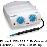
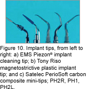
DENTSPLY Professional (Figure 2) Coltene-Whaledent (Figure 3), Parkell (Figure 4), Tony Riso Co, USI, and Odontoson all make magnetostrictive units and tips. Tips are also available from Hu-Friedy, Discus Dental, and Premier Dental. The only magnetostrictive implant tips that can be safely used on titanium implants are the blue plastic tips from Tony Riso Co (Figure 5).
The Parkell Turbo-Sensor or Clean Machine accepts magnetostrictive tips from different manufacturers. Parkell makes the Burnett Power Tip, which is the only thin magnetostrictive tip specially designed for use on high power (Figure 4). All metal, thin tips are also made by most manufacturers and can also be used on medium to high power. Higher power is necessary when removing hard calculus from roots in deep periodontal pockets.
Piezoelectric
Piezoelectric ultrasonic units are used widely in Europe and Asia. The power is generated from a series of ceramic discs or quartz plates that create a linear tip motion without producing heat. Piezoelectric units offer a wide selection of tips that are screwed into the handpiece and tightened with a wrench. The EMS MiniPiezon (Figure 6), Satelec P5 Newtron (Figure 7) , Brasseler NSK Varios 350 (Figure 8) and the Vista Piezo Advantage (Figure 9) all offer compact, table top units with a large selection of tips. Piezoelectric tip designs for these units include large tips for heavy calculus removal, curet tips for light calculus removal, thin tips for biofilm or calculus removal, and diamond coated tips for removal of fine or burnished calculus. Satelec also makes plastic carbon composite implant tips that are used on low power (Figure 10). 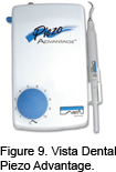 Piezoelectric units are also available from Pro-Dentec, Parkell, and Amdent. Unlike most magnetostrictive thin tips, piezoelectric thin tips can be used with higher power on hard calculus in deep periodontal pockets. The Brasseler NSK Varios 650 (Figure 7) is the only small piezo unit that features a fiber optic light in the handpiece.
 Satelec, Brasseler NSK, and Vista Dental make curet tips and very thin diamond coated tips that are used on low power for removal of thin burnished or embedded calculus (Figure 11). These tips can be used in furcations, root depressions, and CEJ areas where calculus is often burnished and difficult to remove with conventional ultrasonic tips. Large diamond coated tips are very efficient in calculus removal1,2 but are limited to use only during periodontal surgery,where they can be closely observed and controlled. Very thin diamond tips are not as dangerous as large diamond tips when used with light strokes on low power.3 If a clinician attempts to turn up the power with these very fragile thin diamond tips, they will break before they can cause damage to the root surface. Observation with the dental endoscope shows that stroking repeatedly in the same spot for a long time or using the point of a diamond coated tip can result in excess removal of root structure and scratching of restorative materials. Therefore, it is best to use a limited number of strokes with diamond tips or, ideally, they should be used while viewing simultaneously with the dental endoscope.
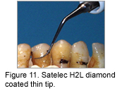
Research studies comparing magnetostrictive and piezoelectric ultrasonic scaling have shown that both methods are equally effective in calculus removal.4,5 Overall, the research is not conclusive on whether one type of ultrasonic is better than another. One study showed that the piezoelectric scaler was more efficient than the magnetostrictive scaler in removing calculus but left the instrumented tooth surface rougher.6 However, a previous study that compared the effect on root surfaces of three ultrasonic scalers to a periodontal curet showed that the curet produced the smoothest surface and the two piezoelectric scalers both produced smoother tooth surfaces than the magnetostrictive scaler.7
CRA Associates, an independent product evaluation service, has rated both magnetostrictive and piezoelectric units very highly in past evaluations.8 The effectiveness of any ultrasonic unit and its tips is determined by the clinician’s experience, technique, and the type of patient being treated. Therefore, the question of whether one type of ultrasonic scaler is better than another is a matter of personal preference that can only be determined after the clinician has had an opportunity to use the ultrasonic scaler, preferably on a number of patients with various degrees of periodontal disease.
The table that accompanies this article can be found here.
References
- Vastardis S, Yukna RA, Rice DA, Mercante D. Root surface removal and resultant surface texture with diamond-coated ultrasonic inserts: an in vitro and SEM study. J Clin Periodontol. 2005;32:467-473.
- Yukna RA, Scott JB, Aichelmann-Reidy ME, LeBlanc DM, Mayer ET. Clinical evaluation of the speed and effectiveness of subgingival calculus removal on single-rooted teeth with diamond-coated ultrasonic tips. J Periodontol. 1997;68:436-442.
- Drisko CL, Cochran DL, Blieden T, et al. Position paper: sonic and ultrasonic scalers in periodontics. Research, Science and Therapy Committee of the American Academy of Periodontology. J Periodontol. 2000;71:1792-1801.
- Jotikasthira NE, Lie T, Leknes KN. Comparative in vitro studies of sonic, ultrasonic and reciprocating scaling instruments. J Clin Periodontol. 1992;19:560-569.
- Busslinger A, Lampe K, Beuchat M, Lehmann B. A comparative in vitro study of a magnetostrictive and a piezoelectric ultrasonic scaling instrument. J Clin Periodontol. 2001;28:642-649.
- Cross-Poline GN, Stach DJ, Newman SM. Effects of curet and ultrasonics on root surfaces. Am J Dent. 1995;8:131-133.
- CRA Associates. Automated Scaler Comparison (Comparison of 16 Ultrasonics and 7 Sonic Scalers). Available at: www.cranews.com/additional_study/1998/98-06/scalers/. Accessed October 13, 2005.
From Dimensions of Dental Hygiene. October 2005;3(10):28, 30-31.
