Early Detection of Gingival Cancer
While rare, gingival cancer can be deadly, and patients’ first line of defense is in the hands of their oral health professional. Frequently mistaken for periodontal disease or local tissue trauma, gingival cancer is thought to arise from a preexisting leukoplakia or erythroplakia found on the gingiva. Are you prepared to catch the signs and symptoms of gingival cancer?
Photo Credit: Sasha wildpixel/ iStock / Getty Images Plus

Age
The average age of the patient at initial presentation of gingival squamous cell carcinoma (SCC) is between 60 and 70.
Photo Credit: Jacob Wackerhausen / E+

Low Risk
SCC often develops in individuals considered at low risk for oral cancer, including nonsmokers and individuals who consume little or no alcohol.
Photo Credit: Aquir / iStock / Getty Images Plus
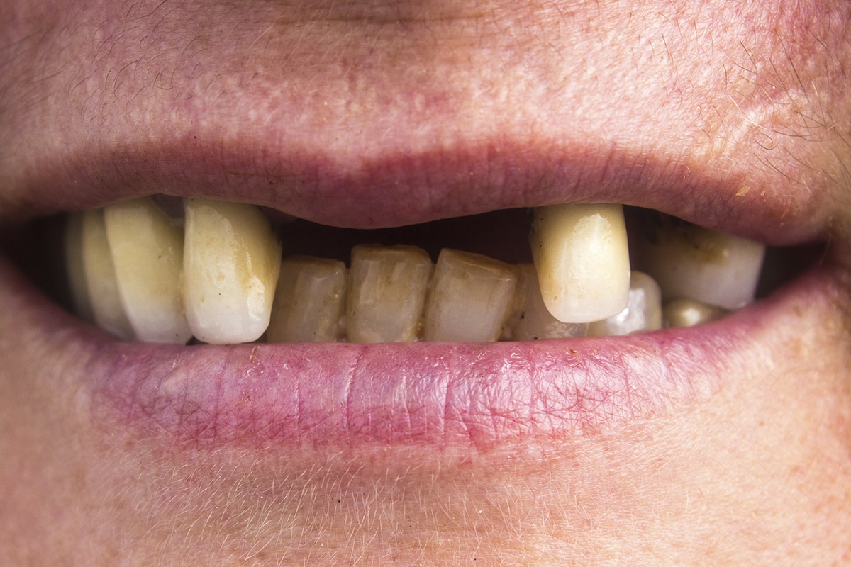
Signs and Symptoms
The signs and symptoms of SCC include red/white lesions, ulceration, loose teeth, swelling, pain, or a nonhealing extraction socket, but these may all be indicative of another oral health problem, making early detection challenging.
Photo Credit: Ismailciydem/ iStock / Getty Images Plus

Lesions
Tumors at more advanced stages will present with a mass or lower lip paresthesia, especially if the inferior alveolar nerve is involved. As a rule, any gingival lesion presenting with the previously described signs and symptoms that has not resolved within 3 weeks to 4 weeks should be biopsied.
Photo Credit: zuper_electracat / iStock / Getty Images Plus
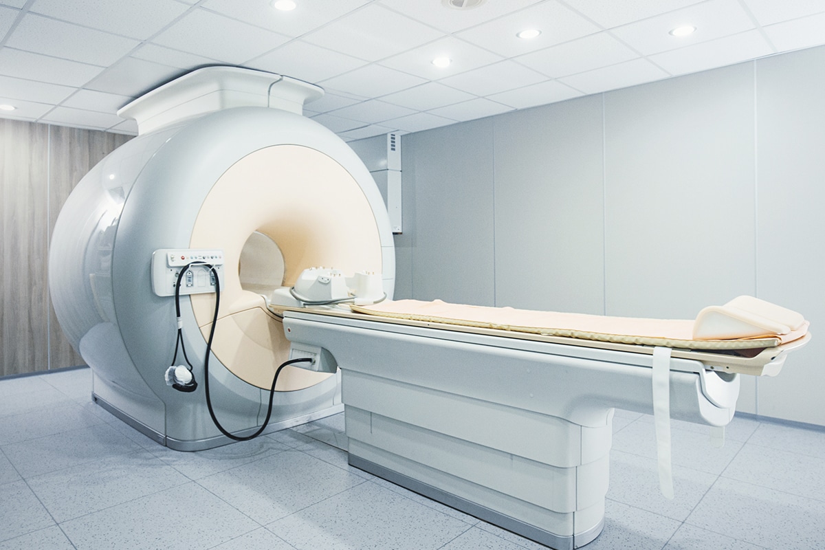
Imaging
Computed tomography and magnetic resonance imaging are considered first-line imaging modalities in the preoperative management of gingival SCC.
Photo Credit: gilaxia / E+

Tumor Staging
Initial tumor staging requires a thorough clinical and radiographic examination. Tumors are typically staged using the designated tumor, node, metastasis American Joint Committee on Cancer staging system.13 Difficulty with this classification occurs in relationship to advanced size (T4) and the specific gingival subsite.
Photo Credit: maclifethai / iStock / Getty Images Plus
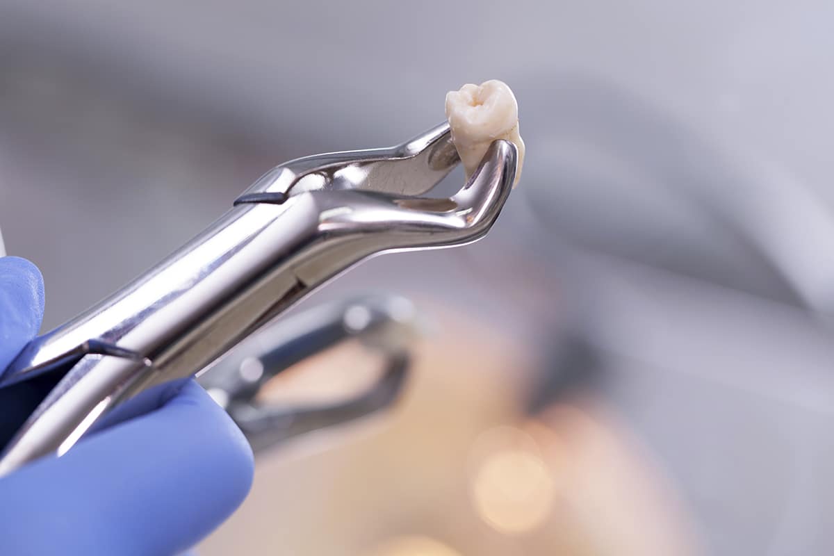
Extractions
If a loose tooth is identified within a gingival mass, it is best not to extract the associated tooth, as this will allow the treating surgeon to assess not only for true bone invasion, but also evaluate the extent of the tumor in the surrounding soft tissue.
