 EDWARDOLIVE/ISTOCK/GETTY IMAGES PLUS; LIGHTHAUNTER/ISTOCK/GETTY IMAGES PLUS
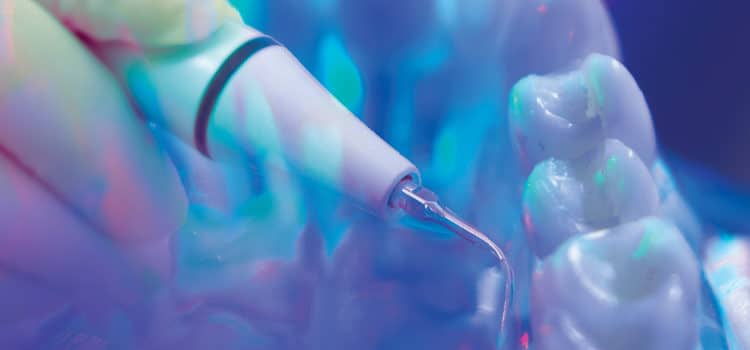
Ultrasonic Instrumentation Tips for Premolars
Effective ultrasonic instrumentation for these teeth depends on the clinician’s knowledge of tooth anatomy, ultrasonic insert/tip designs, and technique.
 Mechanized instrumentation for premolar teeth involves appropriate ultrasonic insert and/or tip (UIT) selection and knowledge about crown and root anatomy. Also, “technique intelligence,” or the combination of concentration, mental imagery, correct technique, and experience, should be considered for proper adaptation and activation of the UIT.Because of the anatomy of premolar teeth, multiple tip designs must be selected to adapt to the curvature, concavities, and grooves (see “Instrumenting Premolars” in the November 2017 issue of Dimensions of Dental Hygiene for figures depicting the typical anatomy of premolars).1A set of straight, right-curved, and left-curved designs is necessary. However, additional inserts with varying shank diameters and tip lengths will also be needed. The size and tenaciousness of the calculus deposit are key to selecting the appropriate UIT diameter, as tip length is relative to probing depth readings. For example, a long, ultrathin UIT is essential for plaque biofilm and fine deposit removal within deep pockets with tight gingival tissue.
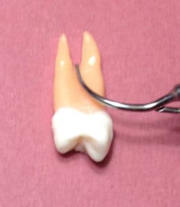
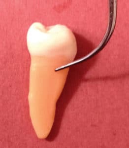
The maxillary first premolar has a significant concavity on the mesial surface, extending from the crown above the cemenotenamel junction (CEJ) to the furcation between the two roots. Additionally, both the mesial and distal surfaces may have furcation involvement with additional concavities. In both of these situations, the insert tip must be adapted to the deposit within the concave surface. Alternatives to accomplishing this adaptation include using the sides (Figure 1) or back of a curved tip on a magnetostrictive insert or the lateral surfaces of a piezoelectric curved tip. A straight tip might be difficult to adapt correctly due to the anatomical curvature of the concavities and furcation roof.
ACTIVE TIP AREA
Using the appropriate active tip area is essential. The portion of the sides or back near the point is most effective for instrumentation. For magnetostrictive technology, the active tip is about 4 mm of the working end, near the point. For piezoelectric unit, the most active area is about 2 mm to 3.5 mm, depending on tip design. Adapting more than the active tip area can cause burnishing, incomplete deposit removal, and inefficient instrumentation. On the other hand, the use of only 1 mm of the tip is likely to produce undesirable root surface alterations. Therefore, the active tip needs to be placed within the root anatomy and should intersect the deposit at different orientations. Use of the UIT’s point is not recommended because of the striations it could cause in the root surface. However, if heavy tenacious calculus is present, then the deposit can be fractured with the point from the top of the calculus down to the epithelial attachment. In the next stage of instrumentation, the UIT is placed near the epithelial attachment to progress upward. Horizontal or oblique orientation of the UIT is particularly useful on the premolar proximal surfaces and might be the best way to reach the midline of the proximal surfaces.
ANGLE
The angle of the active tip to the root should be maintained at 0° to 15°. Because of the crown and root curvatures of premolars, 0° is nearly impossible to achieve and a 15° angle is more likely. This 15° tooth-to-tip angle is ideal because research shows that angulation greater than 15° results in loss of root substance.2,3 In fact, as the tip-to-root angle increases, so does the removal of root substance.2,3
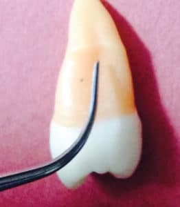
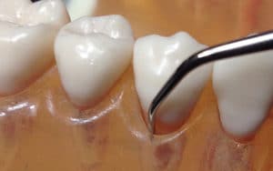
The single-rooted maxillary second premolar has less proximal anatomy than the first maxillary premolar, except in the apical one-third of the root where concavities and grooves are located. A straight UIT will likely be indicated because of the depth of this proximal anatomy (Figure 2, page 31). If significant recession is present, allowing access, a curved UIT might be adapted in the proximal concavities.
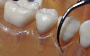
Mandibular first premolars are unique because they have horizontal concavities on both proximal surfaces. They are located below the CEJ to about half way down the root. For these areas, right or left curved UITs can be adapted (Figure 3, page 31). Again, instrumenting at horizontal and oblique orientations might be advantageous in reaching deposits, particularly those located near the midline. Also, the apical portion of the root has concavities with a groove on both the mesial and distal surfaces. If possible, the curved UIT would be ideal to negotiate these concavities; however, because of the depth of this anatomy, a straight UIT might be needed.
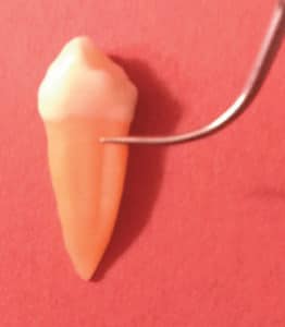
The single-rooted mandibular second premolar has slight root concavities on both the mesial and distal surfaces of the root. These root concavities are more subtle than the concavities on the maxillary first premolar, which create ideal shallow niches for deposit accumulation. Applying the active tip at about 15° at an oblique orientation is probably ideal for this instrumentation (Figure 4).
Typically, the rounded buccal and lingual surfaces of premolars can be instrumented with curved UITs. The easiest strategy is to adapt the curved tip to these surfaces just as a universal curet would be adapted. In other words, on the mandibular right buccal surfaces, use the curved tip that is adapted with the point toward the mesial surfaces. Some clinicians can adapt the curved designs exactly opposite of this concept (Figure 5 and Figure 6). Any time a narrow periodontal pocket is encountered on the buccal or lingual surfaces surface of the premolars, a straight UIT design is indicated with a vertical tip orientation.
UNIVERSAL ACTIVATION
The activation of UITs is not dependent on premolar teeth per se. Instead, the method for correct activation of UITs is universal for all teeth. With a sulcus or shallow pocket depth with light or moderate deposits and plaque biofilm, strokes are activated obliquely from the epithelial attachment to the gingival margin. The presence of heavy deposits indicates a need for the “top-down approach” to break up deposit. Then an upward approach from the epithelial attachment to remove the remaining deposits is appropriate. Areas of roughness can be instrumented with a crosshatching method using different orientations such as horizontal, vertical, and oblique.
CONCLUSION
 Effective ultrasonic instrumentation for these teeth depends on the clinician’s knowledge of tooth anatomy, UIT designs, and technique. Use of the active tip, a 15° tip-to-tooth angle, and various tip orientations are critical to success. For anatomy found subgingivally on premolar teeth, instrument selection and activation are dependent on the location of the gingival margin (near CEJ or recessed) and tissue tone. A straight insert will be needed to reach the anatomy if the gingival margin is near the normal height and a deep periodontal pocket exists exposing root concavities. The curved UIT can be adapted in the deep concavities for effective instrumentation should significant recession be evident.
REFERENCES
- Hodges KO. Instrumenting premolars. Dimensions of Dental Hygiene. 2017;15(12)14–16.
- Flemmig TF, Petersilka GJ, Mehl A, Hickel R, Klaiber B. The effect of working parameters on root substance removal using a piezoelectric ultrasonic scaler in vitro. J Clin Periodontol. 1998;25:158–163.
- Flemmig TF, Petersilka GJ, Mehl A, Hickel R, Klaiber B. Working parameters of a magnetostrictive ultrasonic scaler influencing root substance removal in vitro. J Periodontol. 1998;69:547–553.
![#ad #sponsored
The Cavitron® 300 Ultrasonic Scaling System removes 50% more active biofilm compared to hand scaling.1
Ask your Dentsply Sirona rep how to integrate the Cavitron® 300 System into your hygiene workflow.
https://dimensionsofdentalhygiene.com/cavitron
#DentalHygiene #UltrasonicScaling #Cavitron300
*Clinician in REEL is paid for services.
1. When equal strokes are applied, Cavitron® 300 Ultrasonic Scaling removed on average 50% more active biofilm compared to Hand Scaling in an in vitro study. Johnston, W., Ramage, G., Paterson, M., Brown, J. L., MacKenzie, D., & Culshaw, S. (2022). Effects of instrumentation on in vitro periodontitis biofilm [E-poster presentation]. International Association for Dental Research (IADR). Journal of Clinical Periodontology, 49(Suppl. 23), 143–288. https://doi.org/10.1111/jcpe.13636 Available at: https://iadr.abstractarchives.com/abstract/20iags-3323688/effects-of-instrumentation-on-in-vitro-periodontitis-biofilm. For more information, contact: Consumables-Data-Requests@dentsplysirona.com](https://dimensionsofdentalhygiene.com/wp-content/plugins/instagram-feed/img/placeholder.png)