Transforming Tissues With Phenotype Modification Therapy
This cutting-edge approach redefines the art and science of soft tissue management.
Soft tissue phenotype or gingival phenotype modification aims to enhance esthetics and support periodontal health and stability. Defined as the three-dimensional gingival volume, the term “soft tissue phenotype” was recently adopted by the American Academy of Periodontology as a replacement for “biotype.”1,2
The clinical differentiation between thin and thick soft tissue phenotypes commonly involves visualizing the outline of the probe through the gingival margin.3 Thin phenotype is characterized by tissue thickness less than 0.6 mm, while thick tissue falls within the range of 0.7 mm to 1.2 mm.3 Although the threshold for minimum keratinized tissue width (KTW) remains debated, studies have reported that at least 2 mm of KTW and 1 mm of attached gingiva are crucial for maintaining gingival health and preventing gingival recession.4
Thin soft tissue phenotype has been associated with developing gingival recession and future attachment loss.5 Soft tissue phenotype modification utilizing autogenous grafts (free gingival grafts [FGG], subepithelial connective tissue grafts), or substitutes (acellular dermal matrix or collagen matrices) increases the amount of gingival thickness.6
Soft tissue phenotype modification procedures not only address esthetic concerns but may also re-establish an adequate band of attached and keratinized gingiva that can facilitate the maintenance of oral hygiene.7 This case report presents clinical outcomes of autogenous graft in a patient presenting with gingival recession and thin tissue phenotype in the anterior mandible.
Clinical Presentation
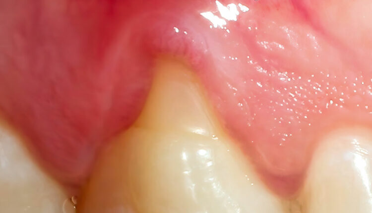
A 45-year-old woman complained of discomfort and receding gums in the front area of her lower jaw. The patient had no relevant medical history. Intraoral examination revealed fair oral hygiene, presence of multiple adjacent gingival recessions, and lack of attached keratinized gingiva at teeth #22, 23, 24, 26, 27, and 28 (Figure 1). The patient was primarily concerned about sites #23 and 24 and provided history of surgical treatment in this area with a nonautogenous soft tissue graft that appeared to have failed due to an infection of the graft .
The gingival recessions were diagnosed as Cairo’s recession type 1 (RT-1) defect. All risks/benefits of the different soft tissue augmentation procedures were discussed. Patient accepted treatment plan for soft tissue phenotype modification using autogenous graft (FGG). Following the initial examination, patient received individualized oral hygiene instructions and professional scaling and polishing 8 weeks prior to surgery.
After profound local anesthesia, the exposed root surfaces were instrumented with Gracey curets and thoroughly rinsed with saline. Mild odontoplasty was completed at site #23 with high-speed instrumentation to remove the soft carious lesion. To prepare the recipient area, a thin partial thickness flap was raised and then excised using horizontal incisions between all teeth at the level of the cementoenamel junction from the distal of #22 to the distal of #27 and two lateral releasing incisions extending well into the alveolar mucosa.

The dimensions of the recipient bed were measured to obtain the autogenous harvest. Partial thickness free gingival grafts consisting of epithelium and a thin layer of underlying connective tissue were harvested from the palate bilaterally adjacent to the premolars and first molars. Harvest sites were covered collagen plugs and sutured with cross sling sutures and the patient was given a thermoplastic palatal stent.
The grafts were trimmed and adapted to the recipient site and stabilized along the coronal and lateral borders with simple interrupted nonresorbable sutures and anchored to the apical periosteum using cross sling resorbable sutures at sites #22, 23, 26, and 27 to ensure compression of the grafts to the recipient site (Figure 2). The two grafts were sutured at the midline with a single simple interrupted suture. After the surgery, the patient received verbal and written postoperative instructions, along with a prescription for 0.12% chlorhexidine rinse. She was advised to refrain from toothbrushing and excessive muscle traction for 2 weeks.
The patient healed uneventfully and appeared to have complied with all the postoperative instructions at the 2-week follow-up, and reported minimal discomfort. All sutures were removed and mechanical plaque control measures were reinstated at this visit. Six weeks postoperatively (Figure 3), the two grafts appeared to have matured and blended. Patient was very satisfied with the outcome as all treated sites presented with significant root coverage and an increase in gingival thickness, vestibular depth, and KTW. Soft tissue phenotype modification procedures allow for improved plaque control, reduced sensitivity, and improved esthetics.5
Barootchi et al6 observed that autogenous soft tissue graft proved superior in all comparisons to graft substitutes for both gingival thickness and keratinized tissue. In this case, the shallow vestibule, thin tissue phenotype, and scar tissue from the prior surgical intervention rendered the site less favorable for gingival augmentation procedures. The successful outcome in this case can be attributed to accurate diagnosis, appropriate treatment selection, and surgical execution.
References
- Jepsen S, Caton JG, Albandar JM, et al. Periodontal manifestationsof systemic diseases and developmental and acquired conditions:consensus report of workgroup 3 of the 2017 World Workshop on the Classification of Periodontal and Peri-Implant Diseases andConditionsJ J Periodontol. 2018;89(Suppl 1):S237-S248.
- Muller HP, Eger T. Gingival phenotypes in young male adults. J Clin Periodontol. 1997;24:65-67
- Rouck T, Eghbali R, Collys K, De Bruyn H, Cosyn J. Thegingival biotype revisited: transparency of the periodontal probethrough the gingival margin as a method to discriminate thin fromthick gingiva. J Clin Periodontol. 2009;36:428-433
- Lang NP, Loe H. The relationship between the width of keratinized gingiva and gingival health. J Periodontol. 1972;43:623-627
- Scheyer ET, Sanz M, Dibart S, et al. Periodontal soft tissuenon-root coverage procedures: a consensus report from the AAP Regeneration Workshop. J Periodontol. 2015;86(2 Suppl):S73–S76.
- Barootchi S, Tavelli L, Zucchelli G, Giannobile WV, Wang HL. Gingival phenotype modification therapies on natural teeth: A network meta-analysis. J Periodontol. 2020;91:1386-1399.
- Pini Prato GP, Franceschi D, Cortellini P, Chambrone L. Long-term evaluation (20 years) of the outcomes of subepithelial connective tissue graft plus coronally advanced flap in the treatment of maxillary single recession-type defects. J Periodontol. 2018;89:1290–1299.
From Dimensions of Dental Hygiene. August/September 2024; 22(5):20-21
