Tips for the Practical Use of Radiography
How to produce high-quality radiographic images while protecting yourself and your patients.
It has been more than a century since radiography was discovered and implemented into health care. Today, dental radiography is a valuable tool in the diagnosis, assessment, and treatment of oral diseases. Dental radiography has experienced an incredible evolution of technology since its inception, but its fundamental concepts remain unchanged. Dental hygienists must continue to be well-versed in the techniques required to generate high quality radiographs while also maintaining important safety protocols.
DENTAL RADIOGRAPHY PROTECTION
The principle of “as low as reasonably achievable” (ALARA) should be followed to ensure the safe use of dental radiography for the operator as well as the patient. The ALARA principle means that every practical protective measure is taken to ensure that the operator and patient will receive the least amount of radiation possible. Collimation, film speed, proper shielding, and digital imaging are also important factors in ensuring safety. Taking only those radiographs necessary for diagnostic and documentation purposes is also aligned with the ALARA principle (see Table 1).1,2
TABLE 1. INDICATIONS FOR RADIOGRAPHY1,2
- Radiographs should be taken in the presence of risk factors for periodontitis or dental caries, and when the health of a tooth is questionable.
- Radiographs should not be scheduled based solely on time, eg, full mouth surveys every 3 years, full bitewings every year.
- Incorporate evidence-based guidelines for the prescription of radiographs created by the American Dental Association and the Food and Drug Administration in 2004. They can be viewed by clicking here.
COLLIMATION
The collimator on the tube head of the X-ray machine is designed to restrict the size of the X-ray beam. Traditional round collimation allows for a small margin of operator error with the diameter of the collimator being 2.75 inches. Consequently, federal law requires the field of radiation at the patient’s skin surface to be no larger than a circumference of 2.75 inches. The diameter of round collimation as mandated by law is nearly three times the area needed to effectively expose a traditional Number 2 intraoral dental film.3
The area in which the patient is exposed can be considerably reduced through further restriction of the X-ray beam by removing the round collimator and replacing it with a rectangular collimator. The absorbed dose of radiation is reduced by 60% to 70% with the use of the rectangular collimator.4 Dental radiographic image quality is also improved because of the restriction of the X-ray beam to the approximate size of the intraoral dental film.
FILM SPEED
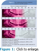
The speed of intraoral dental film is an important aspect of complying with the ALARA principle: the faster the speed, the less radiation exposure time is needed. Figure 1 illustrates the reduction in radiation exposure in relation to film speed as reported by the Food and Drug Administration.5 Using higher speed film complies with the ALARA principle.
Intraoral dental film has emulsion on both sides. The silver halide crystals in the emulsion determine the speed of the film—the larger the silver halide crystals, the faster the film. However, image definition on processed film is more distinctive with smaller silver halide crystals. The larger silver halide crystals used in faster film may result in a grainy appearance. As a result, manufacturers have altered the shape of the silver halide crystals in order to increase the sharpness, resulting in higher contrast images. Clinical trials of the new F-speed film have found that its contrast and resolution are equal to that of D-speed film.6
PROPER SHIELDING
While exposing radiographs, the operator should remain at least 6 feet and 90° to 135° away from the central ray of the X-ray beam. This location will keep the operator out of the direction of the primary X-ray beam. All patients must be properly shielded with a protective apron equipped with a thyroid collar. The use of lead or lightweight alloy aprons in addition to thyroid collars can reduce radiation exposure to the thyroid gland, as well as the ovaries in women and testicles in men, by 94%.7 Lead aprons for patient protection contain lead sheets of at least 0.25 mm, which allow for flexibility and comfort. When lead aprons are not in use, they should be hung up rather than folded. Folding lead aprons will ultimately crack the lead, diminishing its protective capabilities.
DIGITAL IMAGING
Digital imaging is a form of radiography that uses digital X-ray sensors instead of actual film. One of its most significant advantages is the reduction of radiation exposure for both patient and operator. Digital imaging reduces radiation exposure by approximately 90% when compared to D-speed film and 60% when compared to E-speed film.5
Digital imaging systems use sensors to record the penetration of X-ray photons and then transmit the information to a computer, which converts the electronic impulses to a digital image.7 The digital image is composed of pixels. For purposes of this discussion, a pixel is the equivalent of a silver halide crystal in conventional intraoral dental film. When using traditional intraoral dental film, the silver halide crystals are randomly positioned in the emulsion, whereas in digital radiography the pixel has a definite location that can be assigned a number.7 The more pixels in a digital image, the better the resolution and clarity of the final image. See Table 2 for the advantages and disadvantages of digital imaging.3
Indirect digital imaging is a wireless system that uses a photostimulable phosphor plate (PSP) and laser beam scanner to produce an image.7 The PSP plates are not wired and are substantially thinner than wired sensors. The PSP plates store the image on the plate after exposure, which is then placed on a scanner, where the PSP plate is scanned by a laser. After the PSP plate is scanned and the image is viewed on the monitor, the PSP plate must be erased with a high intensity light or the image will remain.
LEGAL ISSUES IN DENTAL RADIOGRAPHY
Dental radiography is essential to assessing and treating patients. Dental radiographs of substandard quality can fail to reveal disease or pathology, compromising patient treatment and putting the oral health care provider at risk of legal consequences in the event of a lawsuit.8
Patient treatment plans are not complete if a full radiographic series is not taken. Radiographs are necessary even in edentulous areas in order to observe retained roots or pathology. Oral health care providers may be held liable if a patient’s record contains an incomplete  radiographic series.8
Different radiographic techniques are used to obtain a full radiographic series on edentulous or partially edentulous patients in order to complete an examination. Panoramic or periapical radiographs of the remaining teeth and edentulous areas are options to complete the radiographic examination. If a panoramic radiograph machine is not available, periapical radiographs of both the maxilla and mandible should be taken in order to meet legal standard of care.8 Dental charts for each patient must contain the correct number of diagnostic-quality radiographs.
CONCLUSION
Dental radiography is an essential component of comprehensive patient care. Radiographs provide an in-depth look at the teeth, supporting structures, disease, and pathology that may otherwise go undetected with a clinical examination. Oral health care providers must be up-to-date about all aspects of dental radiography to be compliant with the standard of care. This working knowledge of dental radiography allows for proper protection of clinicians and patients as well as exposing and producing radiographic images of diagnostic quality.
REFERENCES
- Francisco EM, Horlak DJ, Azevedo S. The balance between safety and efficacy. Dimensions of Dental Hygiene. 2010;8(2):26-30.
- American Dental Association Council on Scientific Affairs. The use of dental radiographs: update and recommendations. J Am Dent Assoc. 2006;137:1304-1312.
- Langland O, Langlais R, Preece J. Principles of Dental Imaging. Baltimore, Md: Lippincott, Williams & Wilkins; 2002:317-319.
- Iannucci J, Howerton L. Dental Radiography: Principles and Techniques. St. Louis: Saunders, Elsevier; 2006:46.
- US Food and Drug Administration. Dental Radiography: Doses and Film Speed. Available at: www.fda.gov/radiation-emittingproducts/radiationsafety/nationwideevaluationofx-raytrendsnext/ucm116524.htm.Accessed January 19, 2011.
- Bernstein DI, Clark SJ, Scheetz JP, Farman AG, Rosenson B. Perceived quality of radiographic images after rapid processing of D- and F-speed direct exposure intraoral x-ray films. Oral Surg Oral Med Oral Pathol Oral Radiol Endod. 2003;96:486-491.
- Frommer H, Stabulas-Savage J. Radiology for the Dental Professional. St. Louis: Elsevier & Mosby; 2005:100,318-321.
- Marsh L. Legal issues in dental hygiene. Access. 2010;24(3):19-21.
From Dimensions of Dental Hygiene. February 2011; 9(2): 52-55.


