The Importance of Probes in Assessing Periodontal Health
Periodontal probes are vital tools in evaluating gingival health, measuring pocket depths, and diagnosing periodontal diseases.
This course was published in the October/November 2024 issue and expires November 2027. The authors have no commercial conflicts of interest to disclose. This 2 credit hour self-study activity is electronically mediated.
AGD Subject Code: 490
EDUCATIONAL OBJECTIVES
After reading this course, the participant should be able to:
- Discuss how the periodontal probe helps support periodontal disease diagnosis.
- Identify the indications for probing and the importance of calibration.
- List advancements in technologies to improve periodontal assessment.
The periodontal probe appears straightforward, but it is also a highly complex instrument. The International Organization for Standardization defines a periodontal probe as a “dental hand instrument designed to measure subgingival pocket depth, used in dentistry for diagnostic purposes and assessment of the condition of periodontal pockets.”1 The probe is used to measure the size of the lesion (extraoral or intraoral) and assess the presence and the extent of periodontal diseases, the distance between teeth (diastema, open bite, overbite, overjet), and detect calculus.2–4
The periodontal probe is integral to evaluating periodontal health.6 Assessment of periodontal conditions using the manual probe is the gold standard for evaluating the adaptation and consistency of the gingiva and its relationship to the tooth.7,8 The probe’s blunt, tapered, circular, or rectangular working end calibrated in millimeter markings makes the insertion safe and measurements easy to read. A working end that meets the shank at an angle of more than 90° allows for better adaptation to all aspects of the tooth.6 Traditional or standard manual probes have colored markings in 1- to 3-mm increments and an average diameter of 0.6 mm.3 These instruments are made of stainless steel, plastic, or titanium.6 The curved Nabers probe is used to evaluate furcation areas and plastic probes are used for dental implants.3 Table 1 provides an overview of the myriad types of periodontal probes.

Role of the Probe in Diagnosing Periodontal Conditions
The key indicators of periodontal health or disease include probing pocket depths, the position of the gingival margin in relationship to the cementoenamel junction (CEJ), clinical attachment loss (CAL), bleeding or suppuration on probing, the width of the attached gingiva, size of oral lesions, and root surface concavities.3,6,7 When utilizing the periodontal probe, two types of depths between the gingiva and the tooth may be measured.7 Biologic depth is the space between the gingival margin and attached tissues. The probing pocket depth is the distance between the base of the pocket and the gingival margin.6 In health, there is a resilient, firm pink attached gingiva with minimal space of periodontal depth.7 The gap or space, called the sulcus in health and the periodontal pocket in disease, can harbor pathogens leading to periodontal destruction and loss of connective tissue.9,10
In the presence of inflammation, the gingiva is flaccid, spongy, loose, and edematous. The accuracy of probing is necessary for identifying and monitoring periodontal diseases. Probing measurements of 3 mm or less are considered healthy; measurements greater than 3 mm may be a sign of disease, inflammation, or gingival overgrowth.7
The biologic width is approximately 2.04 mm in healthy individuals and connective tissue attachment of the dental gingival junction is coronal at the CEJ. CAL is measured from the CEJ to the base of the pocket, indicating true attachment.7,9 Periodontal probes are essential for measuring CAL, periodontal pockets and finding the CEJ.
The presence of bleeding on probing is another indicator of disease. It is best assessed using conventional manual probes and recorded as the proportion of sites with bleeding over all sites evaluated. After an initial record of probing depths, bleeding on probing is visualized about 30 to 60 seconds later.7 Bleeding on probing is described as localized when it occurs in less than 30% of all sites evaluated and generalized when it affects more than 30% of sites evaluated.
In determining the periodontal phenotype, a visual exam and probe are used to estimate the thickness of keratinized gingiva. If a subgingivally inserted probe appears visible through the transparent gingiva, the periodontal phenotype will be established as thin. As this method is not the most reliable in determining thin phenotype, it should be combined with other soft and hard tissue features to confirm thick or thin phenotypes.11
Periodontal probing depths are documented in the patient’s record and used to compare the treated tissues and their response to continued care. Critical depths will be evaluated after initial nonsurgical periodontal therapy to refer for surgery if necessary.12
Indications for Probing
Implants and their attachment differ from natural teeth. There are no periodontal ligaments or gingival fibers, so while probing, the clinician must be careful not to break the biological seal created after placement.13 Aggressive probing can introduce new bacteria and lead to implant failure.14 A plastic probe with a light force of no more than 0.25 N and a diameter of 0.45 mm is recommended.13,14 After 6 months or following osseointegration, implant areas are safe to probe.8,13
Probing depths can evaluate buccal and lingual areas, bleeding, or suppuration to confirm health or disease.13 The sulcus created after an implant has been placed may have a higher probing reading than a natural tooth because of the positioning of the connective tissue fibers. This number acts as a baseline to monitor the implant throughout its existence. Increase in the probing depth could indicate bone loss. Current literature states that metal probes may be used with light force, making them safe for dental implants.14 Plastic probes are still ideal due to their flexibility around the implant.
Assessing the periodontal status of patients undergoing orthodontic care is critical because periodontal diseases usually occur in the age bracket of youths and younger adults.3,15 Gingivitis is a greater risk for orthodontic patients because the appliances can harbor bacteria and biofilm.3,15 Full-mouth probing using a standard periodontal probe should be used during orthodontic treatment and performed every 6 months.16 A diseased pocket will need further evaluation and the patient may be referred to a periodontist, causing a delay or stopping of orthodontic treatment. After orthodontic treatment, periodontal charting should be conducted once a year.15
After completion of nonsurgical periodontal therapy, probing should be done after 4 to 6 weeks when healing has occurred and oral hygiene practices have been resumed.3,14,16 Probing depths are compared with the baseline measurements to assess treatment outcomes.
The Importance of Calibration
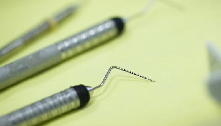
Manual probes are limited in the information they provide about the periodontal pocket.17 Limitations in probing may be related to varying amounts of pressure applied, accuracy in adaptation or angulation during insertion of the probe (Figure 1), reproducibility of the measurement reading, and errors while transferring data to the patient chart.8,17–19 Other errors include insertion of the probe (Figure 2), the diameter of the tip, root concavities, presence of calculus, and inflammation of tissues.3,6,8,9
Assessment can be performed around the deposit or after the removal of calculus to obtain accurate probing depths. Placement errors can interfere with probing due to tooth shape, contact areas, restoration margins, fixed bridges, heavy bleeding, patient difficulty in opening, and macroglossia.3 In advanced periodontal cases with severe inflammation, measuring probing depths after nonsurgical periodontal therapy is more valuable, as it will minimize factors that can prevent obtaining an accurate reading.7

 Varying probing force between examiners can lead to discrepancies in probing measurements: more force will inadvertently lead to a higher measurement. In contrast, an insufficient amount of force can lead to underestimated measurements.20 To mitigate this risk, pressure-calibrated manual probes were developed. While the amount of pressure exerted during probing was calibrated at 20 g, other limitations, such as difficulty cleaning and easy breakage of modified calibrated probes, were noted.18
A new type of handpiece has been developed to precisely measure CAL with the electronic probe.9 A third-generation electronic probe has more controlled force, stores data, creates less discomfort, and causes fewer errors than the original model.9 The device uses Williams’ markings; the measurement is taken and electronically transferred directly into the computer by voice-activated software or a controlled foot switch.2,21 It can also record missing teeth, bleeding, suppuration, furcation, mobility, and plaque. Disadvantages include the size of the probe, difficulty measuring in the proximal surface, and higher cost.5 Clinicians must be trained for proper use; probing depths can sometimes be misinterpreted and lack tactile sensitivity.21
Research on the effectiveness of the electronic probe is mixed. A 2012 and 2013 comparative cross-sectional study showed that manual probes were more reliable and consistent among clinicians than electronic probes.5,10 A 2015 clinical study demonstrated that the electronic probe was more accurate, consistent, and reproducible than a first-generation probe.21
The diameter, size, or shape of the periodontal probe handle or probe itself may cause inaccurate readings. Research has shown that the operator has less hand fatigue, grip force, and tactile precision with a thick-handle probe.10 To compensate, clinicians were observed applying more force when using probes larger in diameter. Ideally, a probe 0.6 mm in diameter should be used with 0.20 g of force to measure periodontal pocket depth accurately.22 Additionally, probes of various thicknesses and different amounts of pressure should be used to evaluate gingival tissues with varying degrees of inflammation.
Discrepancy in probing pocket depths and CAL measurements due to errors in pressure and technique can lead to a misdiagnosis. Manufacturing of the periodontal probe instrument can be off by up to 0.7 mm.6 Therefore, calibration should be increased between multiple clinicians (inter-examiner calibration). This is achieved by using the same type of manual probe within the same clinical setting to avoid misreading the measurement. Frequent clinical exercises are needed to improve calibration in probing technique and the amount of pressure exerted. The goal is to maintain clinically acceptable variance in probing pocket depths and CAL measurement below 1 mm.20
Modern Technologies in Periodontal Assessment
While the use of the manual periodontal probe is still considered the standard of care, emerging technologies are used for calibration purposes, enhancement of findings, improved diagnosis, patient comfort, and to aid in recording and documenting disease progress.
Cone-beam computed tomography (CBCT) is used in diagnosing periodontal diseases but is currently limited to only the evaluation of hard tissues, such as alveolar bone and tooth structures.19 CBCT provides accurate data on furcation involvements and the presence of vertical and horizontal bone loss.23
Ultrasound imaging has great potential in diagnosing periodontal conditions by visualizing all anatomical structures of the periodontium and hard deposits using three-dimensional imaging. The position of the gingival margin, whether coronal or apical to the CEJ, can give incorrect probing depths and is not a true telling of bone destruction. Ultrasonography probing using frequency and computer-aided systems can help identify the gingival sulcus and the actual attachment level.19 Computer-generated algorithms are more consistent and reproducible.19
A 40 MHz periodontal ultrasonography can also assess inflammation, which the operator sometimes overlooks. It can also be repeated to monitor the patient’s status as it does not use radiation or harm the tissue. Ultrasound technology can assess gingival phenotypes, identify early changes in the tissue and lesions, and follow a patient diagnosed with periodontal disease.19
Research shows that ultrasound may be more reliable than manual probing in detecting the position of the gingival margin in relation to the CEJ.24 The 2022 study concluded that ultrasonography is similar in diagnostic effectiveness to traditional probing and also provides more in-depth data on anatomy.24 Limitations to the widespread use of ultrasonography include the equipment size, which creates difficulty in assessing molar areas, the need for clinician training, and cost.
The use of artificial intelligence (AI) can aid in correlating the amount of alveolar bone loss using the information from radiographs and aligning it with periodontal measurements of CAL and probing pocket depths.25 AI can be used to diagnose disease using convolutional neural network algorithms.26 Through periapical and bitewing images, AI can determine the amount of radiographic bone loss and interproximal bone levels. This may minimize operator error and increase accurate diagnoses of periodontal diseases. Oral health professionals’ interpretation depends on their experience and knowledge, thus, the automated assisted program will create more reliable findings.26
Additionally, various applications are used for data input. This allows for voice input of periodontal findings into digital charts with help from AI, complementing conventional probing by decreasing the time needed for charting and analyzing data for case presentation and patient education.
Conclusion
The use of manual periodontal probes continues to be a gold standard in the assessment of periodontal health. New technology developed to improve calibration, ease, and precision in diagnosing periodontal conditions with periodontal probes led to the emergence of pressure-calibrated manual and automatic probes, ultrasonography, CBCT, and the incorporation of artificial intelligence. However, due to the high costs of such equipment and the need for additional clinician training, the use of manual periodontal probes remains widespread.
References
- ISO 21672-1:2012(en)Dentistry — Periodontal probes — Part 1: General requirements https://www.iso.org/obp/ui/en/#iso:std:iso:21672:-1:ed-1:v1:en
- Ko T-J, Byrd KM, Kim SA. The Chairside Periodontal Diagnostic Toolkit: Past, Present, and Future. Diagnostics. 2021; 11(6):932. https://doi.org/葖.3390/diagnostics11060932
- Boyd LD, Mallonee LF, Wyche CJ. Wilkins’ Clinical Practice of the Dental Hygienist. Jones & Bartlett Learning; 2021.
- Rams, Thomas E, and Marc P Manos. “Comparative In Vitro Evaluation of WHO Periodontal Probe and #11/葘Dental Explorer for Subgingival Calculus Detection.” The journal of contemporary dental practice vol. 22,1 13-17. 1 Jan. 2021
- Kour A, Kumar A, Puri K, Khatri M, Bansal M, Gupta G. Comparative evaluation of probing depth and clinical attachment level using a manual probe and Florida proJ . J Indian Soc Periodontol. 2016;20(3):299-306. doi:10.4103/논-124X.181241 https://www.ncbi.nlm.nih.gov/pmc/articles/PMC4976551/
- Gehrig JS, Sroda R, Saccuzzo D. Fundamentals of Periodontal Instrumentation & Advanced Root Instrumentation. Jones & Bartlett Learning; 2019.
- Newman MG, Essex G, Laughter L, Elangovan S. Newman and Carranza’s Clinical Periodontology for the Dental Hygienist. Elsevier; 2020.
- Tankova H, Lazarova Z, Rashkova M. Evaluation of an electronic periodontal probe versus a manual probe in the periodontal diagnosis of children aged 12-14 years. Journal of IMAB – Annual Proceeding (Scientific Papers). 2021;27(4):4087-4091. doi:10.5272/jimab.2021274.4087
- Bansal M, Bareja H, Naveen Kumar P. Comparative assessment of conventional periodontal probes and CEJ handpiece of electronic probes in the diagnosis and primary care of periodontal disease. Journal of Family Medicine and Primary Care. 2021;10(2):692. doi:10.4103/jfmpc_jfmpc_땺_葠
- Van Weringh M, Barendregt D, Rosema N, Timmerman M, Van Der Weijden G. A thin or thick probe handle: Does it make a difference? International Journal of Dental Hygiene. 2006;4(3):140-144. doi:10.1111/j.1601-5037.2006.00187.x
- Abdelhafez RS, Mustafa NM. Determining the periodontal phenotype—probe transparency versus actual: A diagnostic study. Journal of Esthetic and Restorative Dentistry. 2023;35(7):1001-1007. doi:10.1111/jerd.13032
- Caffesse RG, Echeverría JJ. Treatment trends in Periodontics. Periodontology 2000. 2019;79(1):7-14. doi:10.1111/prd.12245
- McCarthy C. Dental implants: to probe or not to probe? Dental Health. January 6, 2016. Accessed January 19, 2024. https://www.swallowdental.co.uk/media/wysiwyg/dental-implants.pdf.
- Gehrig JS, Shin DE, Willmann DE. Foundations of Periodontics for the Dental Hygienist. Wolters Kluwer; 2019.
- Levin L, Einy S, Zigdon H, Aizenbud D, Machtei EE. Guidelines for periodontal care and follow-up during orthodontic treatment in adolescents and young adults. J Appl Oral Sci. 2012;20:399-403.
- Pippi R. Post-surgical clinical monitoring of soft tissue wound healing in periodontal and implant surgery. Int J Med Sci. 2017;14:721-728.
- Moore C, Bai Y, Hariri A, et al. Photoacoustic imaging for Monitoring Periodontal Health: A first human study. Photoacoustics. 2018;12:67-74. doi:10.1016/j.pacs.2018.10.005
- Laugisch, O.; Auschill, T.M.; Heumann, C.; Sculean, A.; Arweiler, N.B. Clinical Evaluation of a New Electronic Periodontal Probe: A Randomized Controlled Clinical Trial. Diagnostics 2022, 12, 42. https://doi.org/葖.3390/diagnostics12010042
- Chifor R, Badea AF, Chifor I, Mitrea D-A, Crisan M, Badea ME. Periodontal evaluation using a non-invasive imaging method (ultrasonography). Medicine and Pharmacy Reports. 2019;92. doi:10.15386/mpr-1521
- Drucker SD, Prieto LE, Kao DWK. Periodontal probing calibration in an academic setting. Journal of Dental Education. 2012;76(11):1466-1473. doi:10.1002/j.0022-0337.2012.76.11.tb05408.x
- Gupta N, Rath SK, Lohra P. Comparative evaluation of periodontal probing depth and attachment levels accuracy using a Florida probe versus traditional probes. Med J Armed Forces India. 2015;71(4):352-358. doi:10.1016/j.mjafi.2012.02.018
- Garnick, J.J. and Silverstein, L. (2000), Periodontal Probing: Probe Tip Diameter. Journal of Periodontology, 71: 96-103. https://doi.org/葖.1902/jop.2000.71.1.96
- Walter, C., Schmidt, J.C., Rinne, C.A. et al. Cone beam computed tomography (CBCT) for diagnosis and treatment planning in periodontology: systematic review update. Clin Oral Invest 24, 2943–2958 (2020). https://doi.org/葖.1007/s00784-020-03326-0
- Moore CA, Law JK, Retout M, et al. High-resolution ultrasonography of gingival biomarkers for periodontal diagnosis in healthy and diseased subjects. Dentomaxillofacial Radiology. 2022;51(6). doi:10.1259/dmfr.20220044
- Kikuchi T, Hayashi J-i, Mitani A. Next-Generation Examination, Diagnosis, and Personalized Medicine in Periodontal Disease. Journal of Personalized Medicine. 2022; 12(10):1743. https://doi.org/葖.3390/jpm12101743
- Chen CC, Wu YF, Aung LM, et al. Automatic recognition of teeth and periodontal bone loss measurement in digital radiographs using deep-learning artificial intelligence. J Dent Sci. 2023;18(3):1301-1309. doi:10.1016/j.jds.2023.03.020
From Dimensions of Dental Hygiene. October/November 2024; 22(6):46-49.
