Support Long-Term Success
Dental hygienists are key to ensuring positive outcomes following periodontal implant therapies.
This course was published in the October 2015 issue and expires October 31, 2018. The author has no commercial conflicts of interest to disclose. This 2 credit hour self-study activity is electronically mediated.
EDUCATIONAL OBJECTIVES
After reading this course, the participant should be able to:
- Discuss the success rates of implant therapy, as well as the prevalence of complications.
- Explain the treatment of peri-implant diseases.
- List the strategies for maintaining peri-implant health.
- Describe the role of the dental hygienist in achieving positive outcomes in implant therapy.
INTRODUCTION
As the number of patients with dental implants increases and dental implant therapy assumes a greater role in the dental practice, clinical investigations have focused on the prevention and management of peri-implant diseases. Clinicians and patients can work together during maintenance appointments to implement evidence-based prevention strategies, one of which is the adjunctive use of chemical antiplaque agents such as a sustained antibacterial toothpaste containing triclosan/copolymer. The early detection and management of peri-implant disease risk factors are key to the long-term success of implant therapy. The Colgate-Palmolive Company is delighted to have provided an unrestricted educational grant to support the article “Support Long-Term Success” in collaboration with the American Academy of Periodontology
—Matilde Hernandez, DDS, MS, MBA
Dental Science Liaison
Colgate Oral Pharmaceuticals
FROM THE AMERICAN ACADEMY OF PERIODONTOLOGY
Dental implant therapies have made healthy, lifelong smiles possible for countless patients. And while these replacement techniques have opened a number of doors, dental implants do not come without complications. In the article “Support Long-Term Success,” American Academy of Periodontology (AAP) member Jonathan H. Do, DDS, educator and California periodontist, addresses the issues that may arise without the proper maintenance and care of an implant. In 2013, the AAP released a report on peri-implant diseases in the Journal of Periodontology, indicating the long-term success of an implant hinges on such factors as treatment planning, execution, and expertise. As specialists trained in the placement of dental implants and in the care of the structures that secure them, periodontists are committed to ensuring patient wellness, reducing complications, and preventing disease. As Dr. Do notes, dental hygienists are also central to the long-term success of implants and positive patient outcomes. As always, the AAP is proud to work with Dimensions of Dental Hygiene and Colgate-Palmolive to provide information that you can apply to daily patient care.
—Joan Otomo-Corgel, DDS, MPH, President, American Academy of Periodontology
Clinical Professor, University of California, Los Angeles School of Dentistry
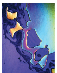
VOLKER STEGER/SCIENCE SOURCE
The introduction of implant therapy, as well as consistent improvement to its armamentarium and technique have changed the landscape of modern dentistry. Prior to osseointegration, clinicians went to great lengths to save compromised teeth. Today, many teeth with questionable or unfavorable prognoses are extracted and replaced with dental implants. Approximately 3 million Americans have implants, and this number is expected to grow by 500,000 per year.1 Mass acceptance of dental implants is due, in part, to their high level of predictability, as well as the various applications for their use—ranging from single tooth to full-arch teeth replacement. While dental implants are an asset to dentistry, no dental replacement can compare to healthy natural dentition. Even with their proven efficacy, predictability, and high rates of success,2–6 complications still arise.
Dental implants are susceptible to mechanical and biological complications. Pjetursson et al2 assessed the 5- and 10-year survival rate of implants and the incidence of technical and biological complications. They reported implant survival rates of 95.6% after 5 years and 93.1% after 10 years. After 5 years, however, biological and mechanical complications were frequent at 33.6%. Two systematic reviews on the 5-year survival rates and incidences of complications of cemented and screw-retained implant reconstructions found both cement- and screw-retained to have comparable survival rates.4,5 Complications, however, increased with the complexity in the implant reconstructions.
Mechanical implant complications frequently involve the implant suprastructure or reconstruction, including ceramic/porcelain fracture and/or chipping, abutment screw loosening, loss of abutment-screw-access-hole restoration, and loss of retention. While the prevalence of abutment screw loosening is less than 5%, it can result in a subgingival gap at the implant-abutment junction that retains plaque similar to an overhanging subgingival crown margin on a natural tooth.2,3
Implant mechanical/technical complications predominantly affect implant restoration, however, biological complications impact the implant fixture and surrounding hard and soft tissue. Inflammatory biological implant complications include peri-implant mucositis and peri-implantitis. Peri-implant mucositis refers to the inflammatory response confined to the soft tissues surrounding the dental implant without the loss of supporting bone beyond the initial bone remodeling during healing.7,8 Clinical signs include mucosal erythema and edema, loose and poorly adapted peri-implant mucosa, marginal bleeding, bleeding on probing, suppuration, tenderness, and probing depths ?4 mm. Radiographically, there is no evidence of crestal bone loss beyond biologic bone remodeling, which can result in crestal bone loss to the implant’s first or second thread.
Peri-implantitis refers to the inflammatory response in the tissue around the implant that has extended beyond the soft tissue to the underlying supporting bone. Bone loss beyond the initial biologic bone remodeling is observed, in addition to the clinical signs associated with peri-implant mucositis. This bone loss can be determined radiographically by comparing geometrically standardized radiographs to the baseline radiograph. If no baseline radiograph is available, a vertical distance of 2 mm from the expected marginal bone level following remodeling post-implant placement is the recommended threshold for the diagnosis of peri-implantitis.9
Due to differences in how peri-implant diseases are defined and diagnosed in various studies, the reported prevalence of peri-implant mucositis and peri-implantitis varies greatly in the literature. A recent systematic review and meta-analysis of 11 studies reported the prevalence of peri-implant mucositis and peri-implantitis ranged from 19% to 65% and 1% to 47%, respectively.10 As such, implants must be evaluated for biofilm-induced inflammatory changes.
Biofilm accumulation has been established as the etiologic factor in the development of peri-implant mucositis.11 The inflammatory process of peri-implant mucositis around an implant is akin to gingivitis around natural teeth, as is peri-implantitis to periodontitis. Peri-implant mucositis is the precursor of peri-implantitis, but peri-implant mucositis does not necessarily progress to peri-implantitis. Similar to periodontitis, peri-implantitis occurs primarily as a result of an overwhelming bacterial insult and subsequent host immune response.8,11 Bacteria associated with periodontitis have been detected at both healthy and diseased implant sites. Diseased sites in patients with periodontitis and sites in smokers have higher concentrations of putative periodontal pathogens such as Tannerella forsythia, Fusobacterium nucleatum, Treponema denticola, and Porphyromonas gingivalis.12,13
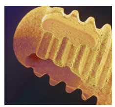
LAGUNA DESIGN/SCIENCE SOURCE
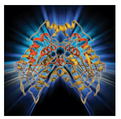
Human tissue biopsies revealed that peri-implantitis and periodontitis lesions have many common features.14,15 Similar markers, such as proinflammatory cytokines, including interleukin (IL)-1?, IL-6, and IL-8 and tumor necrosis factor (TNF)-?, are upregulated in both peri-implantitis and periodontitis (Figure 1).14,16 Assessments of human crevicular fluids also revealed that patients with untreated peri-implantitis contained disproportionately high concentrations of IL-1?, IL-8, TNF-?, and vascular endothelial growth factor.15 A key difference between peri-implantitis and periodontitis, however, is that tissue destruction is more rapid and pronounced in peri-implantitis. In periodontitis, bone loss is usually confined to areas of plaque and calculus accumulation—such as the proximal surfaces, furcations, and root concavities—and manifests as localized vertical infrabony defects. Of course, horizontal bone loss can also occur in periodontitis. In peri-implantitis, bone loss usually happens circumferentially in a pattern called saucerization. The increased susceptibility and pattern of bone loss around implants may be related to the absence of inserting collagen fibers into the implant fixture. With a tooth, the presence of collagen fibers inserting into cementum can confine and separate the inflammatory cell infiltrate and provide a self-limiting process to the inflammatory response. Similar anatomy is absent around an implant.17
TREATING PERI-IMPLANT DISEASES
The outcomes of treating peri-implant diseases are often unpredictable. A review of the literature found that peri-implant mucositis can be effectively treated with nonsurgical mechanical therapy.18 While peri-implant mucositis is reversible at the biomarker level, complete disease resolution is much more difficult to achieve.19 A systematic review20 on the efficacy of patient-administered plaque control to manage peri-implant mucositis and a systematic review21 on the effectiveness of professionally administered plaque removal with or without adjunctive antiseptics or antibiotics in the treatment of peri-implant mucositis found that while disease resolution is possible, it was not achieved in all sites in all patients. Furthermore, the use of adjunctive measures was not found to improve the efficacy of professionally administered plaque removal in reducing clinical signs of inflammation.11,21 Overall, effective patient-administered oral hygiene and professional mechanical debridement can reduce clinical signs of inflammation. Complete disease resolution, however, may not be possible.11,21
The literature also shows that nonsurgical therapy may be ineffective and surgical therapy may be unpredictable in the treatment of peri-implantitis.18 There is also no reliable evidence to suggest which treatment intervention is most effective.22 Implant surface decontamination/disinfection remains challenging, especially for implants with roughened surfaces. Data culled from peri-implantitis patients 1 year or longer after beginning treatment suggested that recurrence of peri-implantitis was high (up to 100%) and retreatment was often necessary.22 Surgical access appears necessary to arrest peri-implant bone loss, and definitive treatment of peri-implantitis may include the removal of the implant, ridge preservation/augmentation, and placement of another implant. Surgical treatments may result in loss of hard and soft tissue and scarring, all of which can compromise esthetics.
Difficulty in treating peri-implantitis may be attributed to several factors, including the absence of connective tissue on the implant fixture surface to help contain infection; the presence of implant threads and roughened implant surfaces, which make surface decontamination challenging, if not impossible; and inadequate hygiene access due to poorly contoured implant suprastructures and lack of vestibular depth. Risk factors for peri-implant diseases include previous periodontal disease, poor plaque control, residual cement, smoking, genetic factors, diabetes, and occlusal overload.8,23,24 The most effective treatment of peri-implant diseases is most likely the prevention of peri-implant mucositis, which can be achieved via effective patient-administered plaque control11 and regular professional maintenance therapy. Early detection and treatment of peri-implant diseases in the peri-implant mucositis phase can also improve the overall prognosis.
OUTCOMES OF PERIODONTAL DISEASE TREATMENT
While gingivitis is ubiquitous and the prevalence of periodontitis among those age 30 and older in the United States is almost 50%, the treatment of chronic periodontitis is relatively predictable when compared to the treatment of peri-implant diseases.25 Gingivitis can be reversed and prevented by patient-administered mechanical plaque removal.26 A systematic review of randomized controlled trials of at least 12 months reported both scaling and root planing and scaling and root planing combined with a flap procedure are effective treatments for chronic periodontitis.27 Results obtained after active nonsurgical and surgical treatment of periodontitis can be maintained long-term with regular professional periodontal maintenance,27–29 even if patient oral hygiene is not ideal.30 The key to long-term periodontal stability is regular professional supportive therapy.29 This provides the opportunity for periodontal evaluation, patient education, and professional mechanical plaque and calculus removal, all of which are essential to maintaining periodontal stability.
PERIODONTAL/PERI-IMPLANT MAINTENANCE
Prevention of biofilm-induced inflammatory periodontal/peri-implant diseases is crucial to the success of long-term maintenance of periodontal and implant therapies. Therefore, periodontal/peri-implant supportive therapy must include thorough evaluation of the periodontal/peri-implant hard and soft tissue, individually tailored oral hygiene instruction, and complete repeated removal of all hard and soft supra- and subgingival deposits.
For both implants and natural dentition, clinical evaluation must include visual assessment of biofilm/calculus accumulation and clinical signs of inflammation—erythema and edema; palpation for suppuration; and evaluation of tissue quality and consistency, marginal bleeding, and mobility. Redness and swelling in the marginal gingiva/mucosa, marginal bleeding, and poor adaptation of soft tissue to the adjacent tooth/implant restoration indicate inflammation in the soft tissue, which is usually caused by biofilm accumulation and inadequate oral hygiene. A tooth is suspended in an alveolus by periodontal ligaments, which inherently allow for physiological movement—a maximum of 0.1 mm to 0.2 mm horizontally and little to no axial mobility. Mobility beyond these levels may indicate either primary or secondary occlusal traumatism. An osseointegrated implant, on the other hand, is in direct contact with bone without any intervening soft tissue, and, therefore, should not exhibit mobility regardless of occlusion, peri-implant inflammation, or bone loss. If an implant exhibits mobility, the suprastructure is loose or the implant has lost osseointegration and is encapsulated in soft tissue.
Probing depths, bleeding on probing, clinical attachment, and furcation invasion around teeth must be evaluated and recorded. While probing ?4 mm around a tooth may indicate disease and pocket formation, probing depths around dental implants do not necessary convey the same implications. Due to the vertical position of the implant and soft tissue thickness, a perfectly healthy implant may have probing depths ?4 mm. In fact, due to the scalloped morphology of the gingival architecture and the flat anatomy of the implant platform, it is common for healthy anterior implants to have proximal probing depths ?4 mm. Furthermore, due to the contour of the implant restoration, obtaining accurate probing depths around an implant can be challenging. An increase in probing depth over time may indicate attachment loss/crestal bone loss in both teeth and implants, thus, changes in probing depth should be monitored. Radiographs of implants taken annually following the baseline radiograph captured at the time of fixture placement can help with the accurate assessment of bone level. Radiographs must be taken with the X-ray beam perpendicular to the implant fixture to allow clear and consistent visualization of the implant platform and threads, which are used as references in the evaluation of crestal bone loss. The lack of tissue resistance to probe penetration and bleeding during probing indicate subgingival pathology in both implants and natural teeth.
Patient education and oral hygiene instruction should follow the periodontal/peri-implant evaluation. Consistent adherence to an individualized oral hygiene regimen is key to achieving gingival health.31 Patients must understand the etiology of periodontal/peri-implant diseases and their responsibilities in disease treatment and prevention. Oral hygiene should focus on biofilm removal along the gingival margins with a toothbrush/ other hygiene aides such as a rubber tip. Patients should be made aware of areas where hygiene is inadequate. They need to understand that plaque removal in these areas can result in bleeding and that thorough hygiene will resolve the inflammation. The use of interdental brushes is one of the most effective tools for interproximal plaque removal around implants and teeth.31,32 In patients unable to effectively remove supragingival biofilms mechanically, the adjunctive use of chemical antiplaque agents, such as triclosan with copolymer, may be required. When used adjunctively to toothbrushing with a fluoride dentifrice, chemical antiplaque agents incorporated into mouthrinse or toothpaste offer significant improvements in managing gingival inflammation and preventing plaque accumulation.32
Professional periodontal/peri-implant maintenance consists of the complete removal of all hard and soft supra- and subgingival deposits. Root surfaces and the transmucosal surfaces of implants should be smooth to minimize plaque accumulation and to facilitate oral hygiene practices.33 Subgingival mechanical instrumentation should be avoided at sites with excellent plaque control and periodontal/peri-implant health to prevent iatrogenic damage to periodontal attachments/implant components. Damage to the periodontal attachments can result in attachment loss and gingival recession in natural teeth. With implants, damage to marginal implant components can create roughened surfaces that contribute to plaque and calculus accumulation and initiate peri-implant diseases. In the presence of plaque, calculus, and tenacious deposits, ultrasonic instrumentation and metal curets can be used to obtain smooth and clean root surfaces. When instrumenting implants, care should be taken to minimize damage to transmucosal implant surfaces. The use of plastic, Teflon-coated, and carbon and gold-coated curets and nonmetal ultrasonic inserts/tips have been advocated to protect titanium implant surfaces and minimize damage. Unfortunately, the large size and flexibility of nonmetal curets may not allow effective plaque and calculus removal.34 The anatomy and physical makeup of implants and their restorations are different from natural teeth. Moreover, curets and scalers were originally designed for scaling and root planing of teeth. Adaptation of scalers and curets to implants are usually poor, and an implant simply cannot be root planed. When used with copious irrigation, ultrasonic instrumentation can help remove implant surface deposits and flush out the peri-implant sulcus/pocket.35 Caution must be exercised to minimize damage, regardless of the instrument used, but the complete removal of all hard and soft supra- and subgingival implant surface deposits is necessary to maintain periodontal health.
THE ROLE OF THE DENTAL HYGIENIST
Dental hygienists are responsible for examining hard and soft tissues for abnormalities; thoroughly removing plaque, calculus, and stain; and properly educating patients about oral health. Patients often spend most of their time during recare visits with dental hygienists, frequently developing a positive rapport. The creation of these trusting patient/provider relationships often encourage patients to ask dental hygienists questions regarding their care as opposed to dentists. Dental hygienists are crucial periodontal care providers in the general dental practice. They are also able to monitor patients over time due to the frequency of recare appointments. As such, dental hygienists act as the first line of defense in protecting patients’ periodontal health. Dental hygienists must be able to recognize symptoms of disease that require treatment and/or referral, including peri-implant diseases, now that implants have become such an integral part of dentistry.
CONCLUSION
Although dental implants are safe and effective, they are not free of complications.2 Much of the literature on implants comes from universities and research centers in which the therapy is provided by highly trained clinicians. An evaluation of complications relating to implants placed in the general practice concluded that implant survival and success rates in general dental practices may be lower than those conducted in academic or specialty settings.6 Considering that more than 3 million implants are placed by general dentists in the US, the number of implants with disease may be staggering.1 With the high prevalence of peri-implant diseases, poor efficacy of peri-implant disease treatments, and the predictability in periodontal disease treatment, preservation of the natural dentition has never been more critical. While it is important for dental hygienists to detect the early signs of peri-implant diseases and educate patients on implant care, their role is even more critical in periodontal maintenance, periodontal disease prevention, and preservation of the natural dentition. The literature consistently shows that after active therapy, periodontal health can be maintained by routine supportive therapy performed by dental hygienists.
ACKNOWLEDGEMENTS
The author would like to thank Kim Thanh Hoang, DDS, for her help with this manuscript; Henry H. Takei, DDS, and his wife June Takei for their unwavering support, mentorship, and friendship; and the American Academy of Periodontology Foundation for supporting his career in academia.
REFERENCES
References
- American Academy of Implant Dentistry. Dental Implants Facts and Figures. Available at:aaid.com/about/press_room/dental_implants_faq.html. Accessed September 8, 2015.
- Pjetursson BE, Thoma D, Jung R, Zwahlen M, Zembic A. A systematic review of the survival andcomplication rates of implant-supported fixed dental prostheses (FDPs) after a mean observationperiod of at least 5 years. Clin Oral Implants Res. 2012;23 (Suppl 6):22–38.
- Wittneben JG, Buser D, Salvi GE, Bürgin W, Hicklin S, Brägger U. Complication and failure rateswith implant-supported fixed dental prostheses and single crowns: a 10-year retrospective study. ClinImplant Dent Relat Res. 2014;16:356–364.
- Wittneben JG, Millen C, Brägger U. Clinical performance of screw- versus cement-retained fixedimplant-supported reconstructions—a systematic review. Int J Oral Maxillofac Implants.2014;29(Suppl):84–98.
- Sailer I, Mühlemann S, Zwahlen M, Hämmerle CH, Schneider D. Cemented and screw-retainedimplant reconstructions: a systematic review of the survival and complication rates. Clin Oral ImplantsRes. 2012;23(Suppl 6):163–201.
- Da Silva JD, Kazimiroff J, Papas A, et al. Outcomes of implants and restorations placed in generaldental practices: a retrospective study by the Practitioners Engaged in Applied Research and Learning(PEARL) Network. J Am Dent Assoc. 2014;145:704–713.
- Lindhe J, Meyle J, Group D of European Workshop on Periodontology. Peri-implant diseases: ConsensusReport of the Sixth European Workshop on Periodontology. J Clin Periodontol. 2008;35(Suppl8):282–285.
- Academy Report: Peri-implant mucositis and peri-implantitis: a current understanding of their diagnosesand clinical implications. J Periodontol. 2013;84:436–443.
- Sanz M, Chapple IL, Working Group 4 of the VIII European Workshop on Periodontology. Clinicalresearch on peri-implant diseases: consensus report of Working Group 4. J Clin Periodontol.2012;39(Suppl 12):202–206.
- Derks J, Tomasi C. Peri-implant health and disease. A systematic review of current epidemiology. JClin Periodontol. 2015;42(Suppl 16):S158–S171.
- Jepsen S, Berglundh T, Genco R, et al. Primary prevention of peri-implantitis: managing periimplantmucositis. J Clin Periodontol. 2015;42(Suppl 16):S152–S157.
- Eick S, Ramseier CA, Rothenberger K, Brägger U, Buser D, Salvi GE. Microbiota at teeth andimplants in partially edentulous patients. A 10-year retrospective study. Clin Oral Implants Res. April 1, 2015. Epub ahead of print.
- Canullo L, Peñarrocha-Oltra D, Covani U, Rossetti PH. Microbiologic and clinical findings ofimplants in healthy condition and with peri-implantitis. Int J Oral Maxillofac Implants. 2015;30:834–842.
- Duarte PM, de Mendonça AC, Máximo MB, Santos VR, Bastos MF, Nociti Júnior FH. Differentialcytokine expressions affect the severity of peri-implant disease. Clin Oral Implants Res.2009;20:514–520.
- Renvert S, Widén C, Persson GR. Cytokine expression in peri-implant crevicular fluid in relation tobacterial presence. J Clin Periodontol. 2015;42:697–702.
- Javed F, Al-Hezaimi K, Salameh Z, Almas K, Romanos GE. Proinflammatory cytokines in the crevicularfluid of patients with peri-implantitis. Cytokine. 2011;53:8–12.
- Berglundh T, Zitzmann NU, Donati M. Are peri-implantitis lesions different from periodontitislesions? J Clin Periodontol. 2011;38(Suppl 11):188–202.
- Renvert S, Roos-Jansåker AM, Claffey N. Non-surgical treatment of peri-implant mucositis andperi-implantitis: a literature review. J Clin Periodontol. 2008;35(8 Suppl):305–315.
- Salvi GE, Aglietta M, Eick S, Sculean A, Lang NP, Ramseier CA. Reversibility of experimental periimplantmucositis compared with experimental gingivitis in humans. Clin Oral Implants Res.2012;23:182–190.
- Salvi GE, Ramseier CA. Efficacy of patient-administered mechanical and/or chemical plaque controlprotocols in the management of peri-implant mucositis. A systematic review. J Clin Periodontol. 2015;42( Suppl 16):S187–S201.
- Schwarz F, Becker K, Sager M. Efficacy of professionally administered plaque removal with orwithout adjunctive measures for the treatment of peri-implant mucositis. A systematic review andmeta-analysis. J Clin Periodontol. 2015;42(Suppl 16):S202–S213.
- Esposito M, Grusovin MG, Worthington HV. Interventions for replacing missing teeth: treatment ofperi-implantitis. Cochrane Database Syst Rev. 2012;1:CD004970.
- Heitz-Mayfield LJ. Peri-implant diseases: diagnosis and risk indicators. J Clin Periodontol.2008;35(Suppl 8):292–304.
- Carcuac O, Jansson L. Peri-implantitis in a specialist clinic of periodontology. Clinical features andrisk indicators. Swed Dent J. 2010;34:53–61.
- Eke PI, Dye BA, Wei L, et al. Update on prevalence of periodontitis in adults in the United States:NHANES 2009 to 2012. J Periodontol. 2015;86:611–622.
- Loe H, Theilade E, Jensen Sb. Experimental gingivitis in man. J Periodontol. 1965;36:177–187.
- Heitz-Mayfield LJ, Trombelli L, Heitz F, Needleman I, Moles D. A systematic review of the effect ofsurgical debridement vs non-surgical debridement for the treatment of chronic periodontitis. Clin Periodontol.2002;29(Suppl 3):92–102.
- Axelsson P, Nyström B, Lindhe J. The long-term effect of a plaque control program on tooth mortality,caries and periodontal disease in adults. Results after 30 years of maintenance. J ClinPeriodontol. 2004;31:749–757.
- Costa FO, Lages EJ, Cota LO, Lorentz TC, Soares RV, Cortelli JR. Tooth loss in individuals underperiodontal maintenance therapy: 5-year prospective study. J Periodontal Res. 2014;49:121–128.
- Ramfjord SP, Morrison EC, Burgett FG, et al. Oral hygiene and maintenance of periodontal support.J Periodontol. 1982;53:26–30.
- Tonetti MS, Eickholz P, Loos BG, et al. Principles in prevention of periodontal diseases: Consensusreport of group 1 of the 11th European Workshop on Periodontology on effective prevention of periodontaland peri-implant diseases. J Clin Periodontol. 2015;42(Suppl 16):S5–S11.
- Chapple IL, Van der Weijden F, et al. Primary prevention of periodontitis: managing gingivitis. JClin Periodontol. 2015;42(Suppl 16):S71–S76.
- Lang NP, Nyman SR. Supportive maintenance care for patients with implants and advanced restorativetherapy. Periodontol 2000. 1994;4:119–126.
- Ramaglia L, di Lauro AE, Morgese F, Squillace A. Profilometric and standard error of the mean analysisof rough implant surfaces treated with different instrumentations. Implant Dent. 2006;15:77–82.
- Roos-Jansåker AM, Almhöjd US, Jansson H. Treatment of peri-implantitis: clinical outcome of chloramineas an adjunctive to non-surgical therapy, a randomized clinical trial. Clin Oral Implants Res. May 26,2015. Epub ahead of print.
From Dimensions of Dental Hygiene. October 2015;13(10):45–50.
