Strategies to Support Patients Undergoing Radiation Therapy
Individuals with head and neck cancer are at high risk of negative oral health effects related to recommended treatment, which frequently includes radiation.
This course was published in the March 2023 issue and expires March 2026. The authors have no commercial conflicts of interest to disclose. This 2 credit hour self-study activity is electronically mediated.
AGD Subject Code: 730
EDUCATIONAL OBJECTIVES
After reading this course, the participant should be able to:
- Explain the common course of radiation treatment and exposure for patients with head and neck cancers.
- List recommended management strategies for this patient population.
- Discuss factors that may increase risk for dental caries, periodontal involvement, and osteoradionecrosis among this cohort.
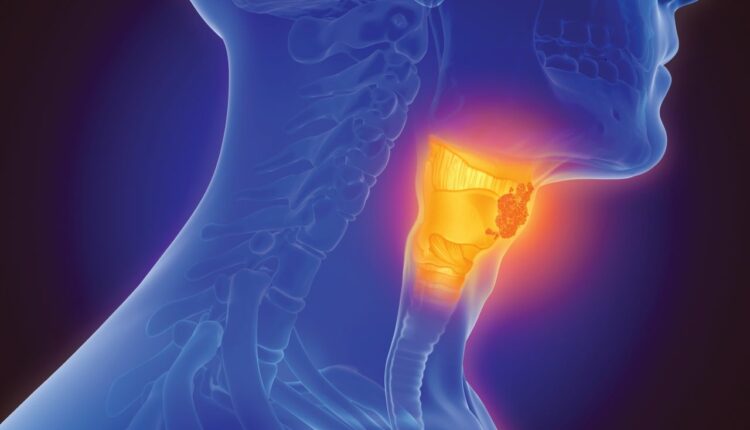
Radiation therapy (RT) is often used for treating head and neck cancers (HNC), including cancers in the oral cavity, oropharynx, larynx, hypopharynx, and salivary glands. The course of RT for such cases usually involves daily fractions of about 2 Gray (Gy), given 5 days per week for 5 weeks to 7 weeks, for a total dose of about 50 Gy to 70 Gy.
This treatment results in several short-term and long-term side effects in the mouth. The short-term side effects can include oral mucositis, reduced salivary flow, candidiasis and taste changes. The long-term effects may include reduced salivary flow and increased risk for dental caries, periodontal diseases, tooth loss, osteoradionecrosis, and trismus.
The OraRad Study
OraRad, a prospective multicenter observational cohort study of 572 patients, is the largest prospective study on oral complications after RT for HNC.1 Participants were evaluated for the study prior to the start of RT and then every 6 months until 2 years after RT.2,3
Of the 572 patients with HNC enrolled in OraRad, 77% were men and the mean age was 58.3 years. Among these subjects, 83% were white, 8% were African American, and 5% were Hispanic. Additionally, 52% were former smokers and 5% were current smokers. Alcohol users comprised 67% of the subject pool, and 64% of the patients reported having dental insurance.
Squamous cell carcinoma had been diagnosed in 82% of patients, salivary gland cancer in 12%, and other types of cancer in the rest. The primary tumor site was the oropharynx, oral cavity in 15%, salivary gland in 10%, larynx/hypopharynx in 7%, and other in the rest.
Therapeutically, 94% received intensity-modulated RT, and 5% received proton therapy. Additionally, 59% of patients had surgery for the HNC, and 68% received chemotherapy.
Salivary Flow
Although most patients received intensity-modulated RT with modern parotid-sparing techniques, stimulated whole salivary flow was greatly reduced at 6 months after RT—to 37% of pre-RT levels. This was followed by a partial recovery in salivary flow, to 59% of pre-RT levels at 18 months.
The average RT dose to the parotid glands was directly associated with the changes in salivary flow after RT. Patients who received less than 20 Gy radiation to the parotid glands had significantly less reduction in salivary flow after RT compared to patients receiving higher doses.4
Patient-reported changes in dry mouth, sticky saliva, swallowing, and mouth opening were significantly worsened at 6 months after RT compared to pre-RT assessments. These quality-of-life assessments were significantly associated with the measured changes in salivary flow. Many quality-of-life indicators demonstrated a partial recovery at 18 months after RT, but were still much worsened compared to pre-RT assessments.
These findings demonstrate there is still a highly significant and persistent reduction in salivary flow and oral health-related quality of life after RT for HNC, even with modern modalities of RT.
Periodontal Diseases
One of the novel findings of this study was that RT led to a striking increase in gingival recession—and in a dose-dependent manner. The mean distance from the cementoenamel junction to the gingival margin worsened significantly during the 2-year study. Greater gingival recession was associated with increased caries on the facial/buccal surfaces.5 These findings help explain the increased risk for cervical caries seen in this population.
The mean clinical attachment loss (CAL) increased modestly from 1.92 mm pre-RT to 2.11 mm at 2 years after RT. The mean periodontal pocket depth slightly decreased from 2.35 mm pre-RT to 2.23 mm at 2 years after RT. The significant apical movement of the gingival margin and only modest increase in CAL resulted in a slight reduction in mean pocket depth.
These findings indicate that oral health professionals can expect patients to have significant gingival recession after RT for HNC. This should be considered during pre- and post-RT dental treatment planning.
Dental Caries
There was a modest increase in dental caries within the first 2 years after RT. The mean score for decayed, missing and filled surfaces (DMFS) was 47.6 at baseline (pre-RT), which increased to 51.9 at the 24-month visit. The increase in DMFS score was significantly smaller for patients compliant with daily prescription-strength topical fluoride use and oral hygiene practices, patients with dental insurance, and those with more than a high school education.6
These findings indicate these patients must use prescription-strength topical fluoride and the importance of maintaining oral hygiene should be highly emphasized. Greater access to dental care and education can also help reduce the caries burden in this population.
Tooth Loss
Tooth loss was defined as a tooth being lost from the mouth or being declared as having hopeless prognosis. Despite comprehensive pre-RT dental management, 17.8% of study patients had a tooth loss event within 2 years of treatment. Risk factors for tooth loss within 2 years included fewer teeth or unrestored caries at the start of RT, greater reduction in salivary flow, and noncompliance with oral hygiene practices.7
The number of teeth at the start of RT is an indicator of prior dental disease experience and also a predictor of future dental disease. Interestingly, the team found that 57% of patients with ≤ 14 teeth at the start of RT lost at least one more tooth during the 2 years after RT compared to only 4% of patients with 28 teeth at baseline (not including third molars).
Patients with unrestored caries at the start of RT were twice as likely to lose a tooth in the following 2 years compared to patients without active caries at baseline. This indicates the importance of restoring active caries as soon as feasible. Logistical issues can make this challenging prior to RT, as the RT usually needs to be started as soon as possible to achieve optimal tumor control.
This suggests that patients with these risk factors should be managed more aggressively during the pre-RT dental management. It should be noted that post-RT dental extractions in areas that have received high doses of RT (usually defined as more than 50 Gy) pose an increased risk for osteoradionecrosis.
Exposed Intraoral Bone/Osteoradionecrosis
Exposed intraoral bone was diagnosed in 35 of the 572 patients (6.1%) in the 2 years after RT. Exposed bone occurred more often in the mandible (76% of episodes).
The mean RT dose to the area of exposed bone was approximately 55 Gy. A diagnosis of osteoradionecrosis was confirmed in 18 patients (3.1%). Of the 35 patients with exposed bone, 24 had a history of a tooth loss event (ie, a tooth extracted or declared hopeless) at or adjacent to the site of exposed bone, either shortly before or after RT. In the other 11 patients, there was no such specific event preceding the exposed bone (nonsurgical spontaneous exposure).8

These findings confirm that osteoradionecrosis is mainly a concern with RT doses > 50 Gy and that dental extractions are a risk factor for exposed bone, but also demonstrate that it can occur without such an event. They likewise reinforce the importance of allowing adequate healing between pre-RT dental extractions and the start of RT (a period of at least two weeks is usually recommended).
Another risk factor for exposed bone was current tobacco use, suggesting an increased level of vigilance in such patients.
Practical Recommendations For Dental Management
The following clinical recommendations are based on the findings of the OraRad study and on other research in the field.9,10
Patients referred for dental evaluation and management prior to HNC RT:
- Consult a radiation oncologist for the radiation field plan for the jaws and planned start date of RT. The parts of the jaws that receive more than 50 Gy will be at higher risk of developing osteoradionecrosis.
- For teeth in areas that will receive more than 50 Gy and have a poor long-term prognosis, consider dental extraction at least 2 weeks prior to the start of RT. Monitor to ensure adequate healing before start of RT. Inform the radiation oncologist if the dental extractions will change the vertical dimension of occlusion, as this impacts the radiation treatment planning.
- Restore active caries before RT if logistically possible; if not, restore as soon as feasible after RT.
- Prescribe prescription-strength topical fluoride (eg, 1.1% sodium fluoride toothpaste).
- Educate the patient on the higher risk for salivary hypofunction, caries, gingival recession, tooth loss, and osteoradionecrosis after RT, and on the importance of maintaining excellent oral hygiene.
- Patients should be informed about the potential for trismus due to radiation fibrosis. Mouth opening exercises should be considered in coordination with their medical providers.
Patients who have received RT for HNC:
- In the 1 month to 2 months immediately following HNC RT, patients are likely to still experience ulcerations of the oral mucosa and have difficulty swallowing and related pain and nutritional compromise. Taste changes can persist for several months after RT.
- Consult the radiation oncologist for the radiation field map for the jaws. The parts of the jaws that have received > 50 Gy are at higher risk of developing osteoradionecrosis.
- Promptly restore caries and manage periodontal disease to avoid the need for extractions in areas that have received more than 50 Gy RT.
- If an extraction becomes necessary in such an area, consider referral to an oral and maxillofacial surgeon.
- For patients with hyposalivation, prescribe high-strength topical fluoride (eg, 1.1% sodium fluoride toothpaste). Recommend strategies to manage hyposalivation and xerostomia, such as sugar-free gum, over-the-counter gels/mouthrinses, and prescription drugs that increase salivary flow (eg, pilocarpine and cevimeline).
- Reinforce to the patient the increased risk for dental disease and the importance of maintaining excellent oral hygiene and receiving routine dental care.
- Emphasize the importance of mouth opening exercises to minimize long-term trismus secondary to radiation fibrosis. Patients with significant trismus should be encouraged to see a physical therapist.
- Screen for oral candidiasis and recurrence of HNC.
Conclusion
Patients receiving RT for HNC are at significant risk for oral disease following RT, including reduced salivary flow, and increased risk for caries, gingival recession, tooth loss, osteoradionecrosis and trismus. The recently published findings of the OraRad study help identify risk factors for adverse dental outcomes after HNC RT and facilitate the evidence-based dental management of this patient population.
ACKNOWLEDGMENT: The authors gratefully acknowledge the contributions of the study patients and study personnel at each site. The OraRad study is funded by the United States National Institute for Dental and Craniofacial Research through a grant (U01 DE022939).
References
- Lalla RV, Long-Simpson L, Hodges JS, et al. Clinical registry of dental outcomes in head and neck cancer patients (OraRad): Rationale, methods, and recruitment considerations. BMC Oral Health. 2017;17:59.
- Brennan MT, Treister N, Sollecito T, et al. Dental disease prior to radiation therapy for head and neck cancer. J Am Dent Assoc. 2017;148:868–877.
- Brennan MT, Treister NS, Sollecito TP, et al. Epidemiologic factors in patients with advanced head and neck cancer treated with radiation therapy. Head Neck. 2021;43:164–172.
- Lin A, Helgeson ES, Treister NS, et al. The impact of head and neck radiotherapy on salivary flow and quality of life: Results of the OraRad study. Oral Oncology. 2022;127:105783.
- Lalla RV, Treister NS, Sollecito TP, et al. Radiation therapy for head and neck cancer leads to gingival recession associated with dental caries. Oral Surg Oral Med Oral Path Oral Radiol. 2022;133:539–546.
- Brennan MT, Treister NS, Sollecito TP, et al. Dental caries post-radiotherapy in head and neck cancer. JDR Clin Trans Res. Epub ahead of print April 11, 2022.
- Brennan MT, Treister NS, Sollecito TP, et al. Tooth failure post-radiotherapy in head and neck cancer. Int J Radiat Oncol Biol Phys. 2022;113:320–330.
- Treister NS, Brennan MT, Sollecito TP, et al. Exposed bone in patients with head and neck cancer treated with radiation therapy: An analysis of the observational study of dental outcomes in head and neck cancer patients (OraRad). Cancer. 2022;128:487–496.
- Levi LE, Lalla RV. Dental treatment planning for the patient with oral cancer. Dent Clin North Am. 2018;62:121–130.
- Sroussi H, Epstein J, Bensadoun RJ, et al. Common oral complications of head and neck radiation therapy. Cancer Med. 2017;6:2918–2931.
From Dimensions of Dental Hygiene. March 2023; 21(3)42-45.


