Radiography provides information that cannot be identified clinically, such as interproximal caries, apical pathologies, or level of alveolar bone loss (Figure 1). The purpose of digital imaging is the same as film radiography but it uses electronic sensors and computer imaging systems that almost instantly produce images on computer monitors instead of film or processing chemicals.1
Three pieces of equipment are needed for digital radiography:
 1. A source of x-radiation. For many offices, the wall-mounted dental x-ray unit is the radiation source, which is compatible with digital imaging as long as the timer can adjust to lower exposure settings.
2. Intraoral sensor. Sensors are small detectors placed intraorally to capture the radiographic image. Initially, sensors were thick, bulky, uncomfortable, and demonstrated surface areas much smaller than traditional film. Currently, sensors are the same size as 0, 1, 2, and 4 size films and are more comfortable. Sensors are either wireless or attached to the computer system by a wired cable.
3. A computer. After the electronic signal is received, a computer converts it from the sensor into the various shades of gray seen on the monitor. This analog picture is divided into squares (pixels) and the computer software takes the information from the analog, gives each pixel a number from 0 to 255, and digitizes the image. The numbers represent a range from black (0) to white (255); all other numbers refer to shades of gray.
Image Acquisition
Currently, two methods exist for acquiring a digital image—indirect and direct. Digital images can be produced indirectly by scanning traditional radiographs using a flatbed scanner with a transparency adapter. The image can then be manipulated using computer software. This method is helpful for dental practices with many patient radiographs mounted in each chart. Although it is time consuming to scan all the radiographs, the images are then converted to digital format and permanently saved in the patient’s electronic file.
The use of photostimulable storage phosphor plates (PSP) is another type of indirect digital imaging. Storage phosphors convert x-rays into light for detection by a receptor, much like the materials used in intensifying screens. Instead of using a sensor connected by a wired cable, this system uses reusable imaging plates coated with phosphors. These plates are slightly flexible and are placed intraorally like a film. After exposure to radiation, the plate is removed from the mouth and placed into an electronic processor where a laser scans the plate and produces an image that is transferred to a computer screen.
The second method of image acquisition is direct digital imaging. The sensor used in direct imaging is a charge coupled device (CCD) or a complementary metal oxide semiconductor (CMOS). The CCD has silicon crystals arranged in a lattice with the ability to convert light energy into an electronic signal. The pixel is the digital equivalent of the silver halide crystals used in film emulsion of conventional radiography, but pixels are not randomly arranged. CMOS detectors use active pixel technology and are less expensive to manufacture. The sensor is exposed to radiation, captures the image, and transfers it to a monitor where the image appears on the screen within seconds.
The CCD contains pixels that sense transmitted light from the radiation and translate it into an electronic message. Since the sensor cannot store information, it must be connected by optic wires to the computer monitor. This electrical lead wire can make this type of imaging bulky and awkward to use. Once the image is produced, it is projected immediately on the computer monitor. This time-saving image production may be beneficial during a difficult dental procedure.
CCD and CMOS sensors render an image almost immediately—no additional processing steps are required. The scanning times of the PSP sensors vary, ranging from 90 seconds to 4 minutes.
Getting the Technology Right
Choosing a digital imaging system is usually based on information from salespeople, articles promoting technology, and various presentations or continuing education courses. In general, dental practices that expose patients to numerous full mouth series (based on patient need) should consider indirect digital imaging with PSP technology. The PSP plates are relatively inexpensive and reusable after sterilization.
 Dental practices that need images immediately should consider direct digital imaging systems. The sensors are more expensive but they produce the images within seconds of exposure. Although digital radiographic imaging systems can be purchased exclusively by themselves, administrative dental software can now incorporate digital systems into one operating system. Well-integrated systems are designed to mesh with the work flow of the dental team and allow providers to concentrate on their main goal—providing exceptional patient care.1
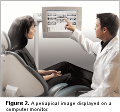
Patient education is an advantage of digital radiography. Viewing the radiographic image on a computer screen provides a visual representation of disease, which is more influential than a verbal explanation accompanied by a small intraoral film. Digital imaging allows patients to become more involved in their treatment choices as it enhances the diagnosis process.2 The more the patient understands, the more likely treatment plans will be accepted. For example, the ability to reveal locations of interproximal calculus and the resultant signs of periodontal destruction may make a greater impression on the patient than discussion alone (Figure 2).
Another major advantage is the reduction of radiation exposure. Radiation exposure is 50% to 80% less than is required for E-speed film used in conventional radiography.1 In keeping with the “as low as reasonably achievable” (ALARA) principle, every possible method of reducing exposure to radiation should be used to minimize risk for the patient and operator.
The digital image can be manipulated to make the radiograph more diagnostic, moving from a darker to lighter image or one with increased contrast. The image can also be magnified or sharpened. These features optimize the image without sacrificing its integrity. Additional image enhancements include zoom features, image orientation, and inverse filtration, which allows the gray scale to be reversed.
Saving Time and the Earth
Time is important in a busy dental practice. With direct digital imaging using a CCD system, the radiographic image is displayed almost immediately after exposure. With a computer monitor at chairside, the operator has the information instantly (Figure 3). Although when using PSP, time is needed to scan the plates.
 Once exposed, digital images can be transmitted instantly to other dental offices or insurance companies. The digital image can be incorporated into the patient electronic record and archived. Multiple copies of the image are a click away, as well as the ability to compare radiographs over time.
In addition, digital imaging is considered environmentally friendly. Since processing chemicals are not used for digital imaging, no disposal issues exist. Many states have established laws to govern the disposal of harmful radiographic processing chemicals.3
Obstacles
Cost is a definite factor when considering a digital imaging system. Depending on the system purchased, the initial cost may be quite high. Reusable PSP plates average approximately $21 to $25 per plate.4 CCD sensors can vary from $5,000 to $7,000 each for an adult size sensor. However, money is saved on the purchase of film, chemicals, space used for storage, the cost of waste disposal, and processor maintenance.
Since sensors come in contact with mucous membrane tissues, they are considered semicritical devices. The Center for Disease Control and Prevention recommends that semicritical devices be cleaned and heat sterilized or cleaned with a high-level disinfectant after use.5 CCD and CMOS sensors cannot be sterilized or autoclaved at this time. These sensors should be protected during use by a Food and Drug Administration-approved barrier, such as a barrier sheath or finger cot, to reduce gross contamination. Disinfecting the sensor with an intermediate level disinfectant is recommended between patient uses. Intraoral imaging sensor manufacturers should be consulted on specific disinfection concerns.
CCD and CMOS sensors may still be bulky for some patients. If proper positioning of the sensor is difficult to achieve, patient discomfort with intraoral projections may lead to a greater number of retakes.6 In addition, PSP plates must be handled very carefully. The sensors scratch easily from fingernails, being dropped on the floor, or contact with debris on counter tops. Operators who attempt to soften the edges may likely find a permanent crease along the surface of the sensor, limiting its diagnostic qualities.
Over the long term, digital imaging systems are a cost effective change from conventional film. Any practice desiring to make the conversion should carefully plan out all details prior to any hardware or software purchases.7
References
- Haring JI, Jansen L. Dental Radiography: Principles and Techniques. 2nd ed. Philadelphia: W.B. Saunders Co; 2000:
- Miles DA, Langlais RP, Parks ET. Digital X-rays are here; why aren’t you using them? J Calif Dent Assoc. 1999;27:926-934.
- Frommer HH. Radiology for Dental Auxiliaries. 7th ed. St Louis: Mosby Inc; 2001.
- Mauriello SM, Platin E. Dental digital radiographic imaging. J Dent Hyg. 2001;75:323-330.
- Kohn WG, Collins AS, Cleveland JL. Guidelines for infection control in dental health-care settings—2003. MMWR Recomm Rep. 2003;52(RR-17):1-61.
- Versteeg CH, Sanderink GC, van Ginkel FC, van der Stelt PF. An evaluation of periapical radiography with a charge-coupled device. Dentomaxillofac Radiol. 1998;27:97-101.
- Schleyer TK, Spallek H, Bartling WC, Corby P. The technologically well-equipped dental office. J Am Dent Assoc. 2003;134:30-40.
From Dimensions of Dental Hygiene. September 2005;3(9):34, 36.
