Instrumentation of Biofilm
A blended approach incorporating both ultrasonics and manual instrumentation best serves the periodontal patient in maintenance.
JW Costerton, PhD, head of the Center for Biofilm Engineering at Montana State University, Bozeman, recently described biofilm as a sophisticated network of well-organized microbes, complete with ventilation, plumbing, nourishment, communication, and modes of travel.1 The deep subgingival environment with its warm, darkened recessed contours must surely serve as “the promised land” for these dynamic congregations. His description of “living veneers” held together by “goo” (extracellular matrix) creates a vivid image of community life along vast microscopic stretches of root surface exposed by attachment loss in the periodontal patient.
These dense aggregations of subgingival microbes harbor periodontal pathogens within their layers. They use defensive strategies and are firmly bound in their surface adherence. Factor in complex root morphology, varying attachment topography plus limited instrument access and subtract the sense of sight, and you have one hefty challenge. What this boils down to in the mechanical removal of periodontal biofilm is that direct physical contact through some form of instrumentation is a requirement for thorough debridement.
Dangerous Assumptions
In maintenance patients with moderate to advanced attachment loss, periodontal debridement can be a time-consuming task that may be underestimated. When patients are compliant with plaque control efforts and on a close interval schedule, they frequently appear well maintained. With limited to no mineralized deposits and isolated bleeding points, debridement procedures can become somewhat hurried (or excessively abbreviated) if assumptions are made about what cannot be seen.
The case for meticulous technique during root debridement may seem less compelling with these patients. However, furcation exposures and scattered 5 mm to 6 mm pockets being maintained nonsurgically are the very conditions that demand 100%. By the time the interval between visits has elapsed, patients have often approached the limit of their biofilm threshold, beyond which the bacterial load is fully capable of overwhelming the host response. Thus, our ability to find and disrupt the farthest reaches of plaque that patients are incapable of accessing becomes critical. It is the primary defense by which they are able to avoid periodontal breakdown.2,3
.jpg)
Photo Courtesy of Hu-Friedy Manufacturing Co.
Periodontal Debridement
Periodontal debridement encompasses all procedures that restore the gingival tissues to health—scaling and root planing (initial therapy) and root debridement (maintenance therapy).
The purpose of root debridement during maintenance is to remove plaque biofilm, endotoxin, and recently formed, lightly mineralized calculus that may have developed between maintenance appointments. The instrumentation to accomplish this objective is primarily a deplaqueing procedure. Root debridement is not intended to remove cementum, dentin, or heavy calculus.4 Instead, it aims to gain access to every square millimeter of subgingival surface in order to mechanically disrupt soft but adherent deposits in a comprehensive manner while also removing any mineralized deposits.
Despite the lack or limit of mineralized deposit, root debridement can be a demanding endeavor in the presence of moderate-to-severe attachment loss. The difficulty of working on accentuated root anatomy is further complicated by limited insertion and access to the base of pockets when the gingiva is no longer edematous. Achieving comprehensive coverage of deep subgingival contours requires a focused technique of closely placed, overlapping strokes that leave no portion untouched. This is true whether manual instruments or power instruments are used.
The method of instrumentation should be chosen by evaluating the attachment topography and root sur-face textures occurring throughout the mouth. In this manner, the treatment approach can be based on the specific needs of each patient, increasing effectiveness and maximizing the therapeutic value of the appointment.
At each maintenance appointment, probe the patient to determine pocket topography and note any changes since the previous visit. Probe carefully with angular adjustments to 45° at all root divergence points to examine for furcation invasions. Display the periodontal probing chart throughout the debridement procedure as a visual reference to the depths and shapes of pockets. This provides a topographical road map to guide instrument selection and technique—especially insertion depth.
Ultrasonic Instrumentation
Just as patients are told that no amount of swishing or spraying will remove their proximal surface plaque—they have to use interdental brushes, toothpicks, and/or floss to mechanically scrape it from the tooth surface—so it is with biofilm and our instruments. Nowhere is this more evident than in the case of ultrasonic instrumentation.
Research has indicated that ultrasonic vibrations create turbulence in the microbial colony through cavitation and what is termed “acoustic streaming.”5 This suggests that the imploding effect on cells can even occur beyond the terminal extent of the vibrating tip.
Some clinicians misinterpret this to mean that passing the ultrasonic tip through the pocket several times with brisk scribbling-movements effectively treats the area through cavitation and turbulence of acoustic streaming. The recently introduced dental endoscope allows clinicians a subgingival view of the root surface at 48x by means of a fiberoptic camera with high intensity illumination attached to a probe-like instrument. This technology removes the traditional guesswork from our therapeutic measures and allows an objective visual means of evaluating instrumentation approaches.6,7 Endoscopic observations have documented numerous islands of undisrupted biofilm and calculus, fully adherent to the root surface, left behind following the aforementioned swift-moving ultrasonic technique.7 This cursory approach—bluntly referred to as “drive-by ultrasonic cleaning”—economizes treatment time at the expense of the patient.
The Hazards
Haste is the first hazard of ultrasonic use. The second is insufficient power. While low power is sufficient for disruption of biofilm, it merely burnishes mineralized deposits. If plaque-retentive hard deposits are present subgingivally as root debridement is performed with thin-tipped ultrasonics on low power, they will be smoothed and polished, rendering them difficult or impossible to feel and remove. Many times this is done unknowingly since the ultrasonic tip offers limited tactile sensitivity. Endoscopic observations have substantiated this occurrence during root preparation.7 Calculus is removed in successive layers until only a thin veneer remains, usually in concavities, furcations, and along the cemento-enamel junction.
Clinicians often begin a debridement procedure using the low powered approach, unaware of the extensive burnishing they are inadvertently causing. Burnished calculus is commonly dismissed as “rough roots” or “clean cementum,” but can still harbor and allow recolonization of pathogenic bacteria.7 This is a frequent cause of deep residual low-grade inflammation detectable only by minute bleeding points upon probing. While it may seem harmless in comparison to the patient’s condition before treatment, this chronic inflammation makes these sites vulnerable to attachment loss.
Technique Recommendations
To remove hard deposits, use heavier ultrasonic tips on medium to high power or use sharp-bladed manual instruments with sufficient lateral pressure. If deposits have become burnished, a periodontal file may be the only instrument that can remove it. The Hirschfeld 3/7 and 5/11 files are recommended for this purpose.7
Because most ultrasonic tips feature a straight profile at their terminal, active end, adaptation to the highly variable, curved contours of the root is problematic.8,9 For this reason, supplement ultrasonic use with localized manual instrumentation for root concavities or pronounced contours. This blended approach will accomplish more thorough debridement of biofilm and establish a biologically acceptable tooth surface compatible with health.
Although seemingly effortless, ultrasonic instrumentation is technique-dependent. Adaptation and activation are both fundamental to success. Care in adapting the terminal 2 mm to 3 mm portion of the tip to the root surface and using confined, overlapping strokes at a moderate to slow pace are essential. Maintaining a steady visual focus on methodical stroke patterns activated in precise channels across each surface will assist in attaining comprehensive root coverage.
Ultrasonic Advantages
Ultrasonic instrumentation does have distinct advantages. Narrow profile ultrasonic tips have attracted a loyal following for good reason. They have the ability to access deep, narrow pockets and furcations with relative ease despite the tight tissues often seen in maintenance patients. These areas have been traditionally inaccessible to standard-sized manual instruments and ultrasonic inserts.9
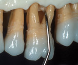
Thin-tipped inserts are especially effective in reducing spirochetes and motile rods in class II and III furcations10 (Figure 1) and should routinely follow the use of mini-bladed manual instruments in these areas.11 Positioning of thin ultrasonic tips featuring a straight terminal portion is identical to probing. The tip is directed apically, while the last 1 mm to 2 mm of the side are in contact with the tooth. Observe care in furcations to prevent perpendicular tip contact, which will gouge the root.
The system and tip should be chosen according to the needs of each patient, but be aware of how differences impact their use. Piezoelectric systems deliver energy to the lateral sides of the tip in a linear motion. Magnetostrictive systems deliver energy to all sides of the tip in an elliptical motion. What the clinician must keep in mind is that only a limited portion of any tip is capable of transferring the maximum energy needed for deposit removal, generally less than 3 mm at the terminal end.
The linear motion of piezoelectric ultrasonic tips is very effective for deposit removal as long as the lateral sides of the tip are kept adapted to tooth surfaces. This requires adjustments of the handle at every line angle by pivoting the fulcrum as instrumentation proceeds around the surfaces of the dentition.
Ultrasonic tip designs continue to evolve with newly emerging profiles that range from roughly the width of an explorer to bladed, beveled, and diamond-coated (Figure 2). Very thin diamond coated ultrasonic tips are able to disrupt and detach biofilm from the root surface by abrasion. Endoscopic observation has also revealed that gentle abrasion with manual diamond files is a new and effective method for fine calculus removal during final root smoothing.7
This capability to directly observe the effects of different systems used at various power levels with an assortment of tips on root surface deposits will continue to enlighten clinicians and stimulate further advances in instrument designs and therapy approaches.
|
Figure 1.Thin magnetostrictive ultrasonic insert in class II/III furcation. |
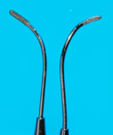
Figure 2. Assorted Satelec piezoelectrictips: H3 Periotip (universal curet), Hi Periotip (diamond tip), #10P, #1 scaler. |
|
|
|
|
Figure 3. Hu-Friedy Mini Five 11/12 curet in buccal furcation. |
Figure4. Brasseler F1 and F2 diamond-coated files. |
.jpg)

Manual Instrumentation
Periodontal maintenance debridement is readily accomplished using extraoral and alternative fulcrums that allow the blade to reach the base of deeper pockets. These are the same handrests often used with ultrasonic instrumentation.
Mini-bladed Gracey curets are recommended for superior adaptation to the accentuated contours of the root. Their small size and rounded backs allow them to be inserted more easily under firm, well-maintained tissue. The short curved blades adapt perfectly into deep proximal concavities, around narrow convexities, depressions of multirooted teeth, and in furcations. These areas are the most vulnerable to breakdown due to the difficulty they pose for patient plaque removal. Therefore, they are the most critical areas to thoroughly instrument (Figure 3).
For maintenance root debridement, the preferred choices are standard Gracey curets and mini-bladed Gracey curets that have been sharpened down to roughly half their width across the face. In this way, the insertion of the instrument in tighter, healthier soft tissue will not be limited.11 Heavier, new rigid mini-bladed Gracey curets should be reserved for initial therapy procedures since blade width and strength are essential for the application of strong lateral pressure during root preparation.
Root debridement strokes are light but overlapping, covering all portions of the root surface. Because they are similar to assessment strokes, the grasp fingers will detect any mineralized deposits or plaque-retentive root irregularities encountered by the blade. If deposits are detected, lateral pressure on the stroke increases to that of a working stroke for deposit removal. The use of sharp curets is imperative for these sensory vibrations to be effectively transmitted to the grasp fingers. Final root smoothing can be accomplished effectively with diamond-coated files that remove biofilm and residual fine calculus (Figure 4).7
Summary
Proficiency in the disruption and removal of periodontal biofilm via root debridement is dependent on accurate assessment (calibrated probing skill); data analysis and treatment decisions, such as the debridement method; instrument/tip selection (size and shape) and technique skill level, such as comprehensive coverage and awareness of root morphology; periodontal probings displayed for reference; and optimally sharpened manual instruments.
An approach that blends manual and powered instrumentation best uses the strengths of each method to benefit the patient in maintenance. The skill and effort to disrupt and thoroughly remove subgingival biofilm will establish and sustain the foundation of periodontal health. Patients can then apply plaque control measures to help maintain the level of health that has been therapeutically established.
References
- Costerton JW, Stewart PS. Battling biofilms. Sci Am. 2001;285(1):74-81.
- Lowenguth RA, Greenstein G. Clinical and microbiological response to nonsurgical mechanical periodontal therapy. Periodontol 2000. 1995;9:14-22.
- Shiloah, J, Patters, M. Repopulation of periodontal pockets by microbial pathogens in the absence of supportive therapy. J Peridontol. 1996;67:130-139.
- Pattison AM. The use of hand instruments in supportive periodontal treatment. Periodontol 2000. 1996;12:71.
- Khambay BS, Walmsley AD. Acoustic microstreaming: detection and measurement around ultrasonic scalers. J Periodontol. 1999;70(6):626-631.
- Stambaugh RV, Myers G, Ebling W, Beckman B, Stambaugh K. Endoscopic visualization of the submarginal gingival dental sulcus and tooth root surfaces. J Periodontol. 2002;73(4):374-382.
- Pattison AM, Pattison GL, Matsuda SA. Periodontal Instrumentation. 3rd ed. Upper Saddle River, NJ: Prentice Hall. In press.
- Drisko CL. Scaling and root planing without overinstrumentation: hand versus power-driven scalers. Curr Opin Periodontol. 1993;3:78.
- Holbrook T, Low S. Power-driven scaling and polishing instruments. Clin Dent. 1989;3:1-23.
- Leon LE, Vogel RI. A comparison of the effectiveness of hand scaling and ultrasonic debridement in furcations as evaluated by differential dark-field microscopy. J Periodontol. 1987;58:86.
- Pattison GL, Pattison AM. Manual instrumentation. In: Newman MG, Takei HH, Carranza F, eds. Carranza’s Clinical Periodontology. 9th ed. Philadelphia: WB Saunders Co; 2002:594-606.
