A Practical Look at Ultrasonic Instrumentation
Stacy A. Matsuda, RDH, BS, shares her expertise on how to maximize your effectiveness when performing power-driven instrumentation.
Q. What is the difference between magnetostrictive and piezoelectric ultrasonic scalers?
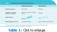
A. Magnetostrictive and piezoelectric scalers move differently. Magnetostrictive scalers use a metal stack to create an elliptical or curved vector of movement at the tip that transfers energy on all sides. Piezoelectric scalers produce linear movement in a flat ellipse initiated by the motion of ceramic rods. For the practicing clinician, an important point is that piezoelectric scalers require precise positioning because their linear motion in and out of the handle means that only the lateral surfaces of the tip transfer the energy necessary for efficient deposit removal. See Table 1 for a comparison of the two different types of power scalers.
THE COMPLEMENTARY APPROACH
Q. Can you describe the complementary approach to instrumentation?
A The complementary approach is based on the idea that neither hand instrumentation nor power instrumentation is a cure-all. Both types of instrumentation have significant strengths that, when used in concert, boost the efficacy of periodontal therapy. Hand instruments have certain access limitations, for instance, reaching the base of deep narrow pockets due to tissue distension and the physical bulk of traditional instruments. Ultrasonic scalers, on the other hand, can reach the base of deep narrow pockets with greater ease using the proper tip. However, ultrasonic tips can sometimes be difficult to adapt to the curvatures, contours, concavities, and longitudinal depressions because almost all ultrasonic tips have a straight edge profile at the point of energy transfer. Hand instruments’ blade curvatures at their terminal third provide a sharp edge to engage calculus and are able to conform to the morphologic contours of root surfaces.
For patients who have pockets or extensive calculus deposits, the complementary approach maximizes efficiency. The approach entails first using the ultrasonic with sufficient power to remove the bulk of calculus deposits, then following with hand instruments to cover root contours, and finally performing a finishing debridement/flush using the ultrasonic scaler with a thin tip. If the patient is healthy and has very little pocketing and/or concave root exposures, hand instrumentation can be limited to essential coverage in proximal contours and furcations. If there are pockets, hand instruments are absolutely necessary as an adjunct. The deeper the probe readings, the more accentuated the contours are, making it exceedingly difficult to gain access. Even with hand instrumentation, specialized hand instruments like minis and files are necessary to reach these difficult areas. For tips on efficacious ultrasonic technique, see the sidebar.
ADVANTAGES
Q. What are some of the benefits of using ultrasonic instrumentation?
A. One benefit to ultrasonic instrumentation is the ergonomic effect. Hand instrumentation can cause severe musculoskeletal strain, fatigue, and pain. Ultrasonic instrumentation is much less taxing on the neuromusculature of the hands. Another significant gain is the ultrasonic’s ability to provide a “super flush” through the action of acoustic streaming and lavage. The ultrasonic scaler can rinse and remove the smear layer (remnants of biofilm) left by hand instrumentation and thus is very effective as the final step prior to dismissal.
Ultrasonic scalers are also perfect to work in class II-III furcations where spirochetes tend to proliferate. These areas are better managed with the skillful use of an ultrasonic. Because root furcations are easily gouged by ultrasonic tips, this type of use is very technique sensitive so clinicians need to be cognizant of the root morphology and careful not to position the tip perpendicular to any surface within the furcation.
COMMON MISCONCEPTIONS
Q. What are some of the more common misconceptions about using power-driven instrumentation?
A. Insufficient power can create major problems that prolong infection. When ultrasonic instrumentation is initiated with thin tips on low power, the clinician can inadvertently polish the outer surface of the mineralized deposits without thoroughly removing them, so they are shaved down until they feel smooth. This smoothness leads the clinician to believe that the surface has been cleaned effectively, but the contaminants within the porous layers beneath the polished outer surface remain. The result is rapid reformation of surface biofilm on the remnants of burnished calculus. Even though a favorable tissue response can be noted from baseline with less overall bleeding, the local infection continues at a lower grade at the point of ulceration.
Another misconception is that ultrasonic scaling is “easy” and “fast.” Ultrasonic instrumentation is easier on the physical body but it is mentally demanding and requires skill. Clinicians must maintain concentration in order to know what parts of the tooth structure have been instrumented. They need to carefully control positioning and movement of the instrument so that the terminal 2 mm of the tip is kept adapted and the fine track of energy transfer has covered subgingival surface areas comprehensively. Because there is no visibility in the pocket, this requires a great deal of focus and visualization of root anatomy. When performed correctly, ultrasonic instrumentation takes an equal amount of time as hand instrumentation.
TIPS FOR EFFICACIOUS ULTRASONIC INSTRUMENTATION TECHNIQUE
- Using the assessment findings, analyze the case type. Determine the efficacy of prior instrumentation and existing care regimen. Remember that bleeding points indicate presence of burnished calculus, which necessitates the use of specialized instruments.
- Formulate a strategy for each patient’s unique case. Where is the disease? What type of deposit is present? What is the degree of mineralization of calculus? How accessible are the deposits? Are there barriers to access (overhangs, furcations, deep vertical defects)? What can be reasonably completed in this appointment?
- Be more conscious of decision making throughout treatment. Apply critical thinking and observe protocols based on current scientific theory.
- Be mindful of attachment topography as you instrument. Post recorded probings in easy view for reference. Probe readings must be taken before instrumentation begins because they provide the road map for instrumentation.
- Maintain awareness of the ultrasonic tip’s pattern of movement across the surface while it is activated. Keep strokes slow, methodical, and comprehensive—not rapid and haphazard. Think about the very narrow track of energy transfer and how precise and overlapping activation strokes must be to cover 100% of the root surface.
- Check the lengths of your tips against a template (available from manufacturers) and replace when they are worn out.
- Have a variety of tips available in your operatory. One tip does not fit all tasks.
- When choosing a tip, consider the profile or cross section of the tip. Tips that are round in shape are more effective for biofilm debridement. The tips that have a beveled, trapezoidal, or rectangular shape in cross section are more effective for calculus removal.
From Dimensions of Dental Hygiene. July 2009; 7(7): 32, 34.


