Implementing Intraoral Scanners
Intraoral scanning devices are used for restorative and preventive care.

Intraoral scanning devices are used for restorative and preventive care. They now have the capability to capture three-dimensional (3D) accuracy of the entire dentition in high resolution, creating records of the hard and soft tissue to monitor oral diseases and conditions. The application of intraoral scanners into every day dental practice enhances the dental and dental hygiene process of care and improves communication between clinician and patient. Digital scans can be easily stored and accessed in the patient’s file and used for interprofessional diagnosis. Chairside intraoral scans allow for immediate viewing of images. CAD/CAM images can be used as a visual aid to improve self-care by demonstrating the health of a patient’s oral cavity.
Photo Credit: alex-mit / iStock / Getty Images Plus
Digital Impressions
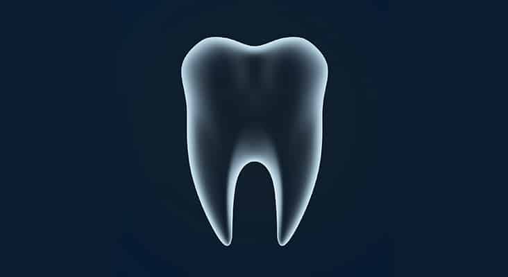
Intraoral scanners capture digital impressions. Comparable to other 3D scanners, intraoral scanners project a light source laser or structured light to scan objects, such as full dental arches, prepared teeth, and implant scanbodies. Intraoral scanners capture images of the hard and soft tissues, as well as restorations. Intraoral images are captured by the scanner and processed by software, which, in turn, generates point clouds. The point clouds are a large collection of points that generate a representation of the existing structures. They are then triangulated by the same software, producing a 3D model of the scanned range. The 3D models of the dentition and tissues are the outcome of the digital impression; they provide an alternative to traditional impression materials and stone models. The intraoral scanner is composed of a handheld camera, computer, and software. The most widely used digital format is the open Standard Tessellation Language (STL) or locked STL-like. This format is a succession of triangulated surfaces in which each triangle is defined by three points and a normal surface. This technique uses a timed laser light directed at the structure and then reflected back to the camera where the data are captured and recorded.
Photo Credit: SuperheroTM / iStock / Getty Images Plus
Parallel Confocal Imaging
Another type of intraoral scanner is parallel confocal imaging. This technique is based on acquisition of focused and defocused images from selected depths. Parallel confocal imaging can detect the sharpness of an area to infer distance to the object that is associated with the focal length of the lens. A tooth can then be reconstructed by consecutive images taken at different focuses and from altered angles around the object, allowing the clinician to directly place the handheld scanner on the tooth and increase stability. The sharpness of the area being scanned is directly related to the dexterity of the clinician, creating distortion and blur if the handheld scanner is not held appropriately.
Photo Credit: Keith Brofsky / Photodisc

Education Possibilities
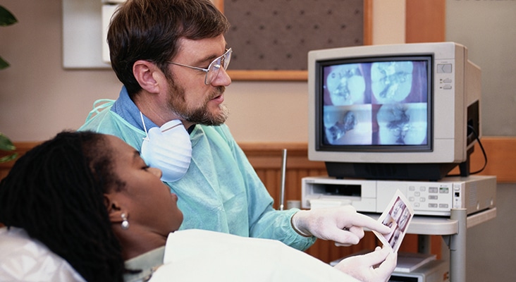
Traditionally, oral hygiene instructions have been provided through videos, printouts, and individual oral communication. Modern technologies, through intraoral images, have changed how oral health professionals can provide patient education. Following a full-mouth intraoral scan, the clinician can use 3D imaging technologies to aid in the detection and diagnosis of dental diseases by providing a magnified, high-resolution image that can be shared digitally with the patient, team members, or specialists, as well as creating a complete patient record. Clinicians with appropriate knowledge and training can complete a full-mouth scan in 3 minutes to 5 minutes. Patients are immediately shown images of fractured teeth, deteriorating restorations, calculus, and gingival inflammation. Oral health professionals can monitor orthodontic relapse, malocclusion, attrition, abfraction, and any changes in gingival recession. Scans can effectively show occlusion and its relevance to oral health. Patients can see areas of heavy occlusal wear and periodontal conditions that may be related to occlusal trauma. Images can also be used to evaluate crowding, overjet, and overbite.
Photo Credit: SDI Productions / E+
Training Is a Must
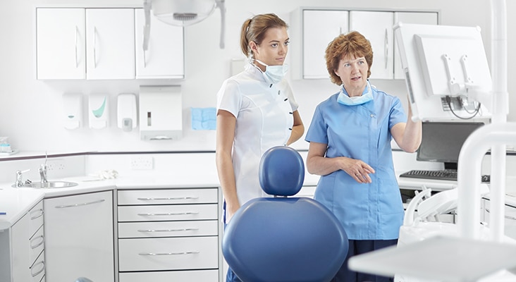
Using the intraoral scanner requires training for all team members. Clinicians with a greater affinity for technology tend to adapt to the new tool more quickly. To better understand the technology and its advantages, offices utilizing intraoral scanners will need to cross-train the dental team. All dental team members need to be comfortable discussing the advantages of the scans used in the office. Knowledgeable clinicians, for example, can describe how CAD/CAM restorations can help retain maximum tooth structure and why this is important to the strength of the tooth and highlight their benefits such as one-visit restorations.
Photo Credit: sturti / E+

Sticker Shock
The initial cost of incorporating intraoral scanning units can be significant. When integrating a digital system into a dental practice, several factors influence cost, including the ability to mill restorations in office. Intraoral scanners can be purchased in open-format, which allows the clinician to immediately open the file once scanned. This format is preferred not only because it is versatile, but because it also reduces future costs incurred in a closed format, such as the need to purchase specific licenses and pay an annual or monthly fee to unlock files. When purchasing open format intraoral scanning devices, a certain degree of experience and knowledge in computers is needed to integrate the system into the existing office workflow. Clinicians who lack computer and software integration skills might opt to purchase a closed format. Closed systems are fully proprietary and do not integrate with other components from other companies.
