 Stock Rojo Verde y Azul / iStock / Getty Images Plus
Cone-Beam Computed Tomography in Implant Therapy
SUBSCRIBE TO THE PERIO UPDATE NEWSLETTER HERE
Cone-beam computed tomography (CBCT) offers three-dimensional (3D) imaging with broad indications for periodontal diagnosis and treatment, as well as all phases of dental implant planning, placement, and follow-up. Yet for all of its advantages, cone-beam imaging should not be used indiscriminately; rather, it should be considered as a powerful adjunct to traditional examinations, diagnosis, treatment planning, and delivery.
The 3D images provided by CBCT offer useful information that can guide clinical decision-making, periodontal diagnosis, treatment planning, and surgical execution (Figure 1). When it comes to diagnosing and managing periodontal diseases, CBCT may be a useful adjunct in certain cases to two-dimensional imaging and clinical probing. Additional periodontal benefits may involve assessing furcation areas or intrabony lesions to help determine treatment potential and prognosis. It is also a useful adjunct in patients requiring orthodontic-periodontic interdisciplinary care.
That said, this imaging modality should be considered an adjunctive diagnostic method after a comprehensive periodontal examination is performed. Limitations to the technology include equipment and software variations that impede the standardization of outcome measures. As indicated by the American Academy of Periodontology’s Best Evidence Consensus Panel on CBCT, areas for investigation include accurately quantifying bone density and changes in linear remodeling, minimizing radiation exposure, and optimizing the image resolution through the reduction of radiation artifacts, beam hardening, and scatter. As is the case with most emerging technologies, the body of knowledge is evolving, and solutions are likely on the horizon.
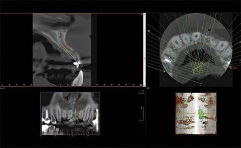
CBCT imaging improves the current standard of care by providing key information, such as measurement of the density, height, and buccolingual width of the alveolar bone at any jaw location, as well as visualization of the pathology, inclination of the bone, and vital anatomic structures. It also aids evaluation of root morphology and pathology at extraction sites.
Using special software, data generated with CBCT imaging during implant planning can be used to fabricate 3D surgical guides. Implants are placed with higher accuracy using these 3D surgical guides. The guides can also be used to generate retro-engineered casts that enable the prefabrication of restorations prior to implant surgery. These restorations can be precisely engineered to improve prosthetic outcomes and delivered to the patient on the day of the surgery. Using all available virtual tools, true restoratively driven implant dentistry can be accomplished via image-guided surgery, benefiting both the patient and dental surgeon. The success and survival of dental implants and implant restorations depend on a thorough diagnosis and careful treatment planning. Not using this imaging modality may increase surgical and prosthetic complications.
This information originally appeared in Frantz BJ. 3D imaging in periodontics. Dimensions of Dental Hygiene. 2019;17(11):23–25.
