Who is at Risk?
While the risk of bisphosphonate-related osteonecrosis is small, dental hygienists need to know who is at risk and what to watch for.
Bisphosphonate-related osteonecrosis of the jaw is defined as “a condition characterized by exposure of bone in the maxilla or mandible presenting for more than 8 weeks in a patient who has taken or currently is taking bisphosphonates and who has no history of radiation therapy to the jaws.”1 The first bishosphonate medications to appear on the market were the nitrogen deficient group of drugs, including clodronate (Bonefos®, Bayer Schering Pharma, Berlin, Germany), etidronate (Didronel®, P&G Pharmaceuticals, Cincinnati), and tiludronate (Skelid®, Sanofi-Aventis, Bridgewater, NJ).
The newer bisphosphonates are nitrogen containing drugs that are deposited on the surface of bone and then internalized by osteoclasts. Second generation bisphosphonates include pamidronate sodium (Aredia®, Novartis Oncology, East Hanover, NJ), which is given intravenously; the oral bisphosphonates alendronate (Fosamax®, Merck & Co Inc, Whitehouse Station, NJ), and residronate (Actonel®, P&G Pharmaceuticals); and ibandronate (Boniva®, Roche Pharmaceuticals, Nutley, NJ), which is dosed on a monthly regime. Zoledronic acid (Zometa®, Novartis Oncology) is a third generation bisphosphonate and is given intravenously.
One theory to explain bisphosphonates’ mechanism of action is the drugs’ osteoclast-inhibiting effect. Bisphosphonates target the mevalonate pathway that ultimately leads to the inhibition of osteoclastic activity by apoptosis.2 Apoptosis is a form of cell death in which a programmed sequence of events leads to the elimination of cells without releasing harmful substances into the surrounding area. Living osteoclasts release the cytokines, bone morphogenic protein, and insulin-like growth factors, which stimulate the formation of new osteoblasts and osteocytes. Osteoblasts and osteocytes have a life span of approximately 150 days. Since the osteoclasts have been inhibited by the bisphosphonates, new bone formation does not occur and the mineral matrix of the dead osteoblasts and osteocytes is not resorbed by the osteoclasts. Thus, bone remodeling and turnover cease to occur and the mature bone becomes acellular and necrotic. The bone also becomes avascular because the capillaries are not maintained.2 Another theory to explain the decreased blood supply was proposed by Santini et al, who showed that bisphosphonates significantly decreased circulating levels of vascular endothelial growth factor.3
INDICATIONS FOR BISPHOSPHONATE USE
Many patients are prescribed oral bisphosphonates for the treatment of osteoporosis and osteopenia, due in part to a decrease in estrogen levels in women4 and a decrease in testosterone levels in men.5 They are also prescribed to treat glucocorticoid-induced osteoporosis, which is sometimes called secondary osteoporosis.6 Glucocorticoids include prednisone and prednisolone and are commonly used to control the symptoms of rheumatoid arthritis and systemic lupus erythematosus. In addition, they are used in the treatment of Paget’s disease, a chronic, metabolic disease that involves bone destruction and growth.7
Bisphosphonates are also used in the treatment of malignant bone diseases. Cancer patients with both primary and metastatic bone lesions develop the skeletal complications of pain, pathologic fracture, spinal cord compression, and hypercalcemia, for which bisphosphonates appear to help.8,9 Bone malignancies secrete osteoclast activating factors. Documentation exists for the successful use of bisphosphonates in reducing bone pain and in treating hypercalcemia and skeletal complications in patients with metastatic breast cancer, prostate cancer, or multiple myeloma.8,9
CLINICAL FEATURES
Bisphosphonate-related osteonecrosis (BRON) is most often seen in cancer patients undergoing treatment for metastatic bone disease. BRON may be present but remain asymptomatic for a long period of time. Marx et al2 reported that approximately one third of the lesions were painless. Tooth mobility and ulceration of soft tissue may be present before BRON is clinically detectable.10 However, BRON becomes symptomatic with pain when there is evidence of inflammation or infection of the soft tissue surrounding the exposed, yellow, necrotic appearing bone.10 The bone itself may be asymptomatic and no bleeding is present on probing. Bony sequestra may be present.11 Infection with drainage of the soft tissue as well as extraoral and intraoral sinus tracts may also been seen. The oral symptoms may be accompanied by fever. These symptoms may occur spontaneously or be triggered by dental treatment, such as extraction or surgery at the site.10
Radiographic evaluation is usually negative in early cases of BRON. When radiographic changes are present, there may be widening of the periodontal ligament or mimicking of classical periapical pathology. Radiographic bone loss between the roots of molars may be present. When there is extensive involvement of the bone, the radiographic appearance is one of diffuse mottling and delayed healing of extraction sites.10
WHO IS AT RISK?
The majority of osteonecrosis cases are in cancer patients who have received intravenous bisphosphonates. Approximately 94% of all osteonecrosis cases are due to intravenous use and 6% are linked to oral bisphosphonates.12 The reason for the increased risk is that the doses for cancer patients are much stronger (up to 12 times higher) than those used for osteoporosis.13 Duration of therapy (longer treatment regimes are associated with a greater risk), along with the presence of medical and dental comorbidities, preexisting dental disease, and invasive dental procedures, are also related to the incidence of BRON.14
Certain procedures are more likely to cause osteonecrosis than others. Recent dentoalveolar trauma appears to be the most common risk factor. A review of 110 patients showed that the majority of osteonecrosis cases were related to the extraction of a tooth or teeth (37.8%).2 The second highest incidence (28.6%) was related to patients with existing periodontal disease. Nase and Suzuki reported a case that occurred after periodontal surgery for crown lengthening.14 In fact, Braun and Iacono reported on a case in 2006 of a 62-year-old Caucasian male diagnosed with multiple myeloma in 1997 and treated with stem cell transplant.15 He was being treated with intravenous zolendronate monthly to prevent metastatic bone disease. The patient had generalized severe chronic periodontitis, gross calculus, and inflamed gingiva. Two months after the second scaling and root planing appointment was completed, the patient presented with inflamed papilla between the mandibular left canine and first premolar, and discomfort in the area. A radiograph revealed a radiopacity consistent with a sequestrum, which was removed. Braun and Iacono hypothesized that the bone became necrotic due to bacterial contamination and exposure during the scaling and root planing.
Osteonecrosis can also occur spontaneously. Merigro et al16 reported on four cases of edentulous patients with no signs of trauma on their oral mucosa. In other cases, trauma to the oral tissues or torus is the culprit in initiating osteonecrosis.
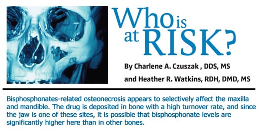
BRON appears to selectively affect the maxilla and mandible. Bisphosphonates are deposited in bone with a high turnover rate, and since the jaw is one of these sites, it is possible that bisphosphonate levels are significantly higher here than in other bones.10 In addition, the oral cavity presents a unique environment. When an open wound is present, such as after an extraction, there is increased risk for compromised healing since there is diminished vascular supply and bacteria present.
CURRENT RECOMMENDATIONS
Once BRON has been diagnosed, treatment consists of preventing progression of the exposed bone with antibacterial mouth rinses and follow-up.17 If the exposed bone is associated with infection and pain, treatment includes pain control, broad-spectrum oral antibiotics, and superficial debridement or removal of bony sequestrum, if present, along with antibacterial mouth rinses.
Although the risk of developing BRON is greatest in cancer patients receiving intravenous bisphosphonates, patients taking oral bisphosphonates for longer than 3 years or patients who have taken oral bisphosphonates for less than 3 years and are also on corticosteroids at the same time may be at a greater risk for developing BRON.17 The estimated incidence of BRON is 0.017% to 0.014%. This rate increases to 0.09% to 0.34% after an extraction. Also, the number of cases has most likely been under reported.17 In order to decrease the risk, it has been proposed that discontinuation of oral bisphosphonates for 3 months before and ater invasive dental surgery may lower the riskof BRON. This must be done in cooperation with the patient’s physician.17
As researchers and clinicians learn more about BRON and who is at risk, better recommendations for prevention will evolve. Presently the American Association of Oral and Maxillofacial Surgeons has developed some management strategies in hopes of minimizing the risk of developing BRON.17 Prior to initiating intravenous bisphosphonate treatment, the patient should have a comprehensive dental examination. Any nonrestorable teeth should be extracted, any invasive dental procedures should be completed, and periodontal health should be optimal. Bisphosphonate treatment should not begin until extraction sites have healed. Placement of dental implants should not be considered in these patients because of the increased chance of bone exposure.2 However, large palatal tori and mandibular tori with thin overlying mucosa should be removed, and antibiotic prophylaxis is recommended for any invasive dental procedure.2
CONCLUSION
As we learn more about osteonecrosis in patients taking this category of drugs and more controlled studies are done, we will be better able to treat it and, more important, prevent it. Patients should be informed of the possibility of this complication occurring and be able to recognize it if it does occur. Intravenous bisphosphonates have been successfully used for more than 15 years in cancer patients with bone metastasis18 and with the increasing use of long-term oral bisphosphonates for osteoporosis, the oral complications of BRON give cause for concern. Emphasis on good oral hygiene and gently supportive periodontal therapy may be extremely important in lessening the risk of BRON. Prospective clinical trials are needed to enable clinicians to make more accurate decisions about risk, preventive measures, and treatment options.
REFERENCES
- Marx RE. Oral and Intravenous Bisphosphonate-Induced Osteonecrosis of the Jaws History, Etiology, Prevention, and Treatment. 1st ed. Hanover Park, Ill: Quintessence Publishing Co Inc; 2007:1, 9-19 .
- Marx RE, Sawatari Y, Fortin M, Broumand V. Bisphosphonate-induced exposed bone (osteonecrosis/osteopetrosis) of the jaws: risk factors, recognition, prevention, and treatment. J Oral Maxillofac Surg. 2005;63:1567-1575.
- Santini D, Vincenzi B, Avvusati G, et al. Pamidronate induces modifications of circulating angiogenetic factors in cancer patients. Clin Cancer Res. 2002;8:1080-1084.
- Kamel HK. Postmenopausal osteoporosis: etiology, current diagnostic strategies, and nonprescription interventions. J Manage Care Pharm. 2006;6(Suppl A):S4-S9.
- Isidori AM, Giannetta E, Pozza C, Bonifacio V, Isidori A. Androgens, cardiovascular disease and osteoporosis. J. Endocrinol Invest. 2005;28(10 Suppl):73-79.
- Compston JE. Emerging consensus on prevention and treatment of glucocorticoid-induced osteoporosis. Curr Rheumatol Rep. 2007;9:78-84.
- Hosking D. Pharmacological therapy of Paget’s and other metabolic bone diseases. Bone. 2006:38(Suppl 2):S3-S7.
- Berenson JR, Lichtenstein A, Porter L, et al. Efficacy of pamidronate in reducing skeletal events in patients with advanced multiple myeloma. N Engl J Med. 1996;334:448-493.
- Hortobagyi GN, Theriualt RL, Porter I, et al. Efficacy of pamidronate in reducing skeletal complications in patients with breast cancer and lytic bone metastasis. N Engl J Med. 1996;335:1785-1792.
- Ruggiero SC, Fantasia J, Carlson E. Bisphosphonate-related osteonecrosis of the jaw: background and guidelines for diagnosis, staging and management. Oral Surg Oral Med Oral Pathol Oral Radiol Endod. 2006;102:433-441.|
- Biasotto M, Chiandussi S, Dore F, et al. Clinical aspects and management of bisphosphonates-associated osteonecrosis of the jaws. Acta Odontologica Scandinavica. 2006;64:348-354.
- Woo SB, Hellstein JW, Kalmar JR. Narrative review: bisphosphonates and osteonecrosis of the jaws. Ann Intern Med. 2006;144:753-761.
- Reid IR, Brown JP, Burckhardt P, et al. Intravenous zoledronic acid in postmenopausal women with low bone mineral density. N Engl J Med. 2002;346:653-661.
- Nase JB, Suzuki JB. Osteonecrosis of the jaw and oral bisphosphonate treatment. J Am Dent Assoc. 2006;137:115-119.
- Braun E, Iacono VJ. Bisphosphonates: case report of nonsurgical periodontal therapy and osteochemonecrosis. Int J Periodontics Restorative Dent. 2006;26:315-319.
- Merigo E, Manfredi M, Meliti M, Corradi D, Vescovi P. Jaw bone necrosis without previous dental extractions associated with the use of bisphosphonates (pamidronate and zoledronate): a four-case report. J Oral Pathol Med. 2005;34:613-617.
- AAOMS Position Paper. American Association of Oral and Maxillofacial Surgeons position paper on bisphosphonate-related osteonecrosis of the jaws. J Oral Maxillofac Surg. 2007;65:369-376.
- Weitzman R, Sauter N, Eriksen EF, et al. Critical review: updated recommendations for the prevention, diagnosis, and treatment of osteonecrosis of the jaw in cancer patients—May 2006. Crit Rev Oncol Hematol. 2007;62:148-15.
From Dimensions of Dental Hygiene. November 2007;5(11): 12-14.

