The Pathway to Synergy
Using complementary instrumentation to achieve the most effective results.
Synergy is defined by Webster’s Dictionary as “a mutually advantageous conjunction of distinct elements.” Applied to therapy, synergism can yield tangible benefits for both the clinician and the patient with increased efficiency, economy of effort, and improved clinical outcomes. A complementary approach to instrumentation where technology and hand skills are blended to accomplish their maximum potential is definitely a synergistic approach.
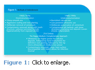
Looking back over the past 5 decades, methods of debridement have varied. At one extreme was aggressive hand instrumentation, often resulting in excess tooth structure removal with pronounced cervical constriction. At the opposite end was the abandonment of hand instruments in favor of low powered ultrasonics used with thin modified tips. Each protocol had its own consequences—unintentional iatrogenic odontoplasty on the one hand and burnished subgingival calculus sustaining periodontal infection on the other (see Figure 1).
The 21st century brings a new model of treatment. While the literature shows that complete calculus removal is not possible, this does not mean that it shouldn’t remain the goal of scaling and root planing (SRP).1-6 The goal of SRP is the formation of a biologically compatible root surface over which the soft tissue can heal, which means thorough removal of root accretions since they are a reservoir for pathogens.7
Biofilm research, along with the introduction of the dental endoscope, has been instrumental in forming a new paradigm for periodontal therapy. Residual calculus can no longer be dismissed as harmless.8-10 A review of the literature comparing hand and ultrasonic instrumentation, while compelling in terms of individual efficacy,11,12 brings up the issue of variables since the debridement process itself is so subjective.
Clinical outcomes are heavily influenced by a host of factors. Were the curets used new and bulky? This may mean that access was compromised. Did the operator possess a strong foundation of instrumentation fundamentals and have experience treating periodontally involved teeth? This may mean that blade adaptation, angulation, adequate lateral pressure, and/or instrument selection was not optimal. Was care taken to comprehensively cover every square millimeter of root surface? The morphologic characteristics of complex root anatomy present a constant challenge that is only compounded by the operator’s relative level of skill and experience.13
THE RATIONALE
To better understand the rationale behind a complementary approach, instrumentation modalities should be closely examined for their limitations. Access is a critical issue in periodontal debridement. Root instrumentation means physical disruption of deposit through direct contact, whether a curet blade is used or an ultrasonic tip. The challenge for both modalities is that ultrasonic tips can reach certain areas less accessible to hand instruments but some specialized hand instruments comform to root curvatures better than ultrasonic tips. In other words, they each compensate for the other’s weakness.
 Adaptation is the conforming of the curet or ultrasonic tip to the tooth. In the presence of periodontal destruction, the broad, flat surfaces of the crown give way to root structures that taper into narrow small radius curvatures, proximal depressions deepen into abrupt concavities, cervical line angles reduce to hairpin-turn convexities, and the three-dimensional obstacle of the furcation further complicates the task. Hand instruments can be adapted anywhere along the working blade, including around the toe. Ultrasonic tips are effective at their maximum peak only at the terminal 2 mm–3 mm. It may seem as though the entire length of the tip is acting on the surface but, in fact, it is a narrow track typically less than 2 mm.
Part of the difficulty in ultrasonic instrumentation is that the curvatures of the tooth do not coincide with the straight profile of the most commonly used ultrasonic tips. This adaptation is better achieved by the curved blade of mini or micro-mini bladed curets. When using the ultrasonic, keep the terminal 2 mm to 3 mm of the tip adapted to the tooth. This maximizes the power transfer at the terminal end and boosts the efficacy of deposit removal.
Once access is gained and adaptation attained, the next essential ingredient is coverage. The way the ultrasonic tip is moved across the root surface affects the success of therapy. The tip needs to contact every square millimeter of subgingival surface. Movement over the tooth should be slow and methodical for complete coverage.
Using the right power settings is key. Without adequate power, mineralized deposits are merely polished over on their outer surface. This burnishing prevents the overlying soft tissue ulceration from healing.14 Once burnished, the deposits are impervious to detection, leading the clinician to believe that they have been eliminated. Compounding the problem is the difficulty of removal. To prevent these problems, use the upper range of power settings (medium high to high power) for calculus deposits.
Following the first round of ultrasonic debridement, horizontal strokes with hand instruments or vertical strokes with mini or micro-mini bladed curets can closely adapt into concave surfaces and effectively cover the deeper contours of root morphology.
Thin tipped ultrasonic inserts on lower power are used as a final step in the process to disrupt remaining biofilm and calculus dislodged by hand instruments. The flushing action of this final step is especially important in furcations where instrumentation is inevitably compromised.
THE BOTTOM LINE
The most important factor for attaining a successful outcome rests in the hands of the clinician. The best instruments and technology will be of little advantage if not used with precision and care. Optimally sharpened hand instruments used with adequate lateral pressure conserve time and effort while reducing the likelihood of repetitive motion injury. Steady awareness maintained to eliminate randomness in ultrasonic stroke activations contribute to an optimized clinical outcome. A complementary approach to instrumentation combines both techniques’ unique advantages to better serve both the patient and the clinician.
REFERENCES
- Sherman PR, Hutchens LH, Jewson LG, Moriarty JM, Greco GW, McFall WT Jr. The effectiveness of subgingival scaling and root planing. I. Clinical detection of residual calculus. J Periodontol. 1990;61:3-8.
- Stambaugh RV, Dragoo M, Smith DM, Carasali L. The limits of subgingival scaling. Int J Periodontics Restorative Dent. 1981;1:31-41.
- Buchannan SA, Robertson PB. Calculus removal by scaling/root planning with and without surgical access. J Periodontol. 1987;58:159-163.
- Fleischer HC, Mellonig JT, Brayer WK, Gray JL, Barnett JD. Scaling and root planing efficacy in multirooted teeth. J Periodontol. 1989;60:402-409.
- Kepic TJ, O’Leary TJ, Kafrawy AH. Total calculus removal: an attainable objective? J Periodontol. 1990;61:16-20.
- Stambaugh RV, McMullin KA. Effectiveness of long term, non-surgical maintenance in deep periodontal pockets. J Dent Res. 1988;67:272.
- Fujikawa K, O’Leary TJ, Kafrawy AH. The effect of retained subgingival calculus on healing after flap surgery. J Periodontol. 1988;59:170-175.
- Chen C, Rich SK. Biofilm basics. Dimensions of Dental Hygiene. 2003;1(1):22-25.
- Stambaugh RV, Myers GC, Watenabe J, Lass C, Stambaugh KA. Clinical response to scaling and root planing aided by the dental endoscope. J Dent Res. 2000;79:abstract 2762.
- Pattison A, Pattison G. Periodontal instrumentation transformed. Dimensions of Dental Hygiene. 2003;1(2):18-20, 22.
- Hallmon WW, Rees TD. Local anti-infective therapy: mechanical and physical approaches. A systematic review. Ann Periodontol. 2003;8:99-114.
- Tunkel J, Heinecke A, Flemmig TF. A systematic review of efficacy of machine-driven and manual subgingival debridement in the treatment of chronic periodontitis. J Clin Periodontol. 2002;29(Suppl):72-81.
- Brayer WK, Mellonig JT, Dunlap RM, Marinak KW, Carson RE. Scaling and root planing effectiveness: the effect of root surface access and operator experience. J Periodontol. 1989:60:67-72.
- Stambaugh RV. Perioscopy—the new paradigm. Dimensions of Dental Hygiene. 2003;1(2):12-13, 15-16.
From Dimensions of Dental Hygiene. July;6(7): 24-26.

