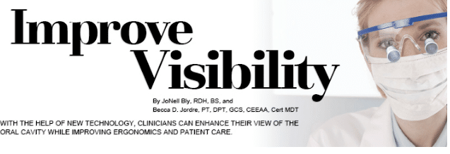
Improve Visibility
With the help of new technology, clinicians can enhance their view of the oral cavity while improving ergonomics and patient care.
Oral health professionals are always looking for ways to improve visibility and enhance ergonomics. Much attention has been given to the problem of musculoskeletal disorders in the dental hygiene community. Results of a 2012 survey of 1,110 practicing dental hygienists indicated that 51% of all participating clinicians had at least one work-related injury.1 The primary injury sites listed were the neck, shoulders, and upper back. These work-related injuries caused 38% of the affected individuals to reduce their work hours permanently.1
Many new products are available to help clinicians establish a healthy work setting. In order to maintain their musculoskeletal health, dental hygienists may want to consider implementing some of these aids into practice. The purpose of this article is to introduce clinicians to products designed to improve visibility in the oral cavity. With increased awareness of these innovations, dental hygienists may be able to refine the ergonomics of their practice and make strides toward a healthy work environment.
For optimal visual acuity, there must be adequate light. Vision is initiated when light is reflected off of objects and enters the eyes.2 The oral cavity is naturally dark and nonreflective; therefore, obtaining sufficient lighting can be challenging. Many clinicians find the traditional overhead lights used in the dental operatory to be insufficient for illuminating the oral cavity. The lack of light often causes clinicians to adopt unhealthy postures for long periods, potentially leading to musculoskeletal problems.
Innovations related to lighting are ongoing, and careful evaluation of these new technologies is essential. For proper eye comfort, the light source must not be on either extreme of the light scale. For example, excessively bright light is perceived as glare and diminishes visual acuity. Incorrect or dim lighting can produce shadowing and also impede vision. When evaluating products designed to improve illumination of the oral cavity, clinicians should consider several factors, including: existing overhead lighting, the reflection of surrounding surfaces, and the amount of natural light present.3
LIGHT-EMITTING DIODE TECHNOLOGY
Dental illumination changed dramatically with the advent of light-emitting diode (LED) technology. These lights produce bright illumination without noticeable radiant heat. Compared to traditional light sources, LED chips are extremely efficient and small, allowing the light and batteries to be attached to the clinician. LED innovation has been incorporated into many dental products with different degrees of success. The intensity of these light systems varies a great deal. Properly designed illumination systems should correctly light the work area and protect the clinician’s eyes from intense light or potentially harmful optical radiation.4 Other sources have stated that it is important to avoid any dental headlight that has too strong of a blue-light component, as this could be potentially hazardous to users.5 Due to these concerns, it is important to purchase dental products from trusted sources.
LOUPES AND LIGHTING
The use of magnification has greatly impacted the practice of dentistry. Magnification provides better visibility in the oral cavity, while also helping clinicians improve their posture. When student posture was assessed using a version of Branson et al’s Posture Assessment Instrument, magnification provided significant posture benefits.6 Other studies have also examined the benefits of magnification in dentistry with similar results.7–9
Many types of loupe frames are available, as well as different magnification levels. To ensure the best fit, clinicians should seek professional help when purchasing loupes. The correct working distance and appropriate declination angle must be determined and measured accurately for the loupe to fit properly. In addition, the frame must correctly fit the clinician’s facial structure for optimal performance. Improperly selected loupes will place the user at an increased risk for musculoskeletal problems.10
The addition of coaxial lighting systems to the lens frame can enhance the value of magnification. When properly mounted on the frame, the light follows the clinician’s eyes—reducing glare and the need to reach to adjust the overhead light during an appointment. Early light systems were heavy and attached to bulky batteries and cords. Recent innovations have reduced the weight of the lights from 30 g in 2009 to less than 10 g in 2012.11 LED dental lights are also available on a head strap or protective frame without magnification and may be useful in certain practice settings. Another advantage of LED lighting is the energy efficiency of the LED chip, which lasts for more than 20,000 hours. These lights are powered by small, portable lithium ion batteries, enabling the clinician to work easily while wearing the light. Several styles of lights are available. Some lenses incorporate the battery pack into the protective frame, eliminating the need for wires.
Vigilant product selection is important when considering purchasing magnification and a headlight. Proper fitting of loupes and headlights by a trained professional is critical to achieving ergonomic excellence. Magnification and lighting systems should be custom fit, and a trial period should be available so the clinician can evaluate the product’s effectiveness. Important factors to consider include the brightness of the light, the ability to easily adjust the brightness, beam uniformity, and weight.12 The LED light must be of adequate intensity and adjustable for various lighting situations, and should provide a neutral white light for clinician safety and proper color accuracy.4
The use of ultrasonic scalers may reduce the stress caused by hand instrumentation and help clinicians prevent repetitive force injuries.13 To enhance the ergonomic benefits, several manufacturers include LED lighting in their ultrasonic scaling units. Lights are available in the handpieces of scalers or in the ultrasonic inserts/tips (UIT). In both cases, the lights are activated when the UIT is in use. These lights greatly improve visibility when performing ultrasonic therapy.14
LED TECHNOLOGY AND RETRACTION AIDS
LED lighting is also available in retraction aids. The use of retraction aids enables the solo clinician to better utilize indirect vision and improve visibility in the oral cavity.15 LED lights are available in retraction and suction devices. This system combines hands-free vacuum suction with intraoral illumination. The light intensity is fully adjustable for brightness and can be found in multiple sizes to fit most patients.
ADDITIONAL AIDS
A simple strategy to utilizing existing operatory lighting is purchasing high-definition mouth mirrors. Several different technologies are employed by manufacturers to create mirrors that provide sharper images and better color visibility. The best options come in variable sizes and have ergonomically designed handles. The addition of anti-fog solutions to the face of the mirror may further enhance this clarity. Previously, dental companies have introduced mouth mirrors with an LED light inserted in the mirror head. Several styles of these LED mirrors are available for home use but none are designed for dental professionals. Mirrors are also available that continually remove water and debris from the mirror face by vibratory action. These mirrors are expensive, which may limit their use at this time; however, the future of this technology is promising.
Intraoral cameras can also enhance visibility of the teeth and surrounding tissue. Images and videos captured with intraoral cameras can be used for patient education. New high-definition intraoral cameras also allow clinicians to visualize oral conditions on computer monitors at a magnification level greater than what can be achieved with dental loupes. Light-induced fluorescence evaluators are also available within intraoral cameras to assist in the detection of gingival inflammation, plaque, and calculus.16 This innovation may enable clinicians to view all areas of the oral cavity in great detail with increased efficiency.
Another advancement for improving operator visibility is endoscopic technology. A periodontal endoscope allows the clinician to obtain a view inside the periodontal pocket. A recent study demonstrated the improvement in subgingival calculus removal during instrumentation when an endoscope was used compared to tactile evaluation only.17
CONCLUSION
Addressing ergonomic challenges in the practice of dental hygiene is an ongoing pursuit. Current technological advances can play a role in this pursuit and may assist in the prevention of work-related stress and injury.
REFERENCES
- Guignon A, Purdy C. Dental Hygiene Practice Survey. Available at: ergosonics.com/OldSite/Student_Handouts_-_2013_files/Students%20-%202013%20presentation%20%20part%20one.pdf. Accessed December 20, 2014.
- Dragoi V. Chapter 14: Visual Processing: Eye and Retina. Neuroscience Online. Available at: neuroscience.uth.tmc.edu/s2/chapter14.html. December 20, 2014.
- Valachi BS. Practicing Dentistry Pain-Free: Evidence Based Ergonomic Strategies to Prevent and Extend Your Career. Portland, Oregon: Posturedontics Press; 2008.
- Ayoub HM, Darby ML. Ergonomic best practices. Dimensions of Dental Hygiene. 2013;11(4):30–34.
- Stamatacos C, Harrison JL. The possible ocular hazards of LED dental illumination applications. J Tenn Dent Assoc. 2013;93:25–29.
- Maillet JP, Millar AM, Burke JM, Maillet, MA, Maillet WA, Neish NR. Effect of magnification loupes on dental hygiene student posture. J Dent Educ. 2008;72:33–44.
- Branson BG, Bray KK, Gadbury-Amyot C, et al. Effect of magnification lenses on student operator posture. J Dent Educ. 2004;68:384–389.
- Maggio MP, Villegas H, Blatz MB. The effect of magnification loupes on the performance of preclinical dental students. Quintessence Int. 2001;42:45–55.
- Sanders MJ, Branson BG, Black MA, Simmer-Beck M. Changes in posture; a case study of a dental hygienist’s use of magnification loupes. Work. 2010;35:467–476.
- Sunell S, Rucker L. Surgical magnification in dental hygiene practice. Int J Dent Hyg. 2004;2:26–35.
- Christensen GJ. Smaller and lighter LED headlamps. Clinician’s Report. 20125(4):1–6.
- Brame J. Seating, positioning, and lighting. Dimensions of Dental Hygiene. 2008;6(9):36–37.
- Ryan DL, Darby JM, Bauman D, Tolle SL, Naik D. Effects of ultrasonic scaling and hand activated scaling on tactile sensitivity in dental hygiene students. J Dent Hyg. 2005;79:9.
- Brame JL. Advances in ultrasonics. Dimensions of Dental Hygiene. 2010;8(10):48–57.
- Valachi BS. Little things can make a difference. Dent Today. 2010;29:140–143.
- Obrochta JC. Efficient and effective use of the intraoral camera. DentalCare.com. Available at: media.dental care.com/media/en-US/ education/ce367/ce367.pdf. Accessed December 20, 2014.
- Osborn JB, Lenton PA, Lunos SA, Blue CM. Endoscopic vs tactile evaluation of subgingival calculus. J Dent Hyg. 2014;88:229–236.
From Dimensions of Dental Hygiene. January 2015;13(1):18,21–23.

