
Extraoral Fulcrums
The essentials of using extraoral fulcrums for periodontal instrumentation.

The teaching and use of extraoral fulcrums is a controversial topic among dental hygiene educators. However, many experienced clinicians use extraoral fulcrums routinely and believe they are essential for advanced periodontal instrumentation, especially in the maxillary posterior areas. Intraoral fulcrums are adequate for supragingival or shallow subgingival instrumentation in these regions, but they present severe limitations for gaining access and establishing proper working angulation when treating deep pockets.
Selecting an Intraoral or Extraoral Fulcrum
A stable intraoral fulcrum is dependent on the relationship between the location of the finger rest and the working area. First, the intraoral finger rest must be located to allow correct positioning of the lower shank to the tooth and, hence, optimal working angulation. At proper working angulation, the lower shank of a Gracey curet is parallel to the tooth surface and the lower shank of a universal curet is tilted slightly toward the tooth.
Second, the intraoral finger rest must be positioned to enable the use of wrist motion. It should be close enough to the working area to allow the middle and fourth finger to be kept together in a “built-up” fulcrum. In any segment of the mouth, as instrumentation proceeds from surface to surface and from tooth to tooth, the location of the intraoral finger rest must be frequently adjusted or repositioned to maintain working angulation and to allow wrist motion. If adjustment or repositioning does not provide a fulcrum that meets these requirements, an extraoral hand rest should be considered.

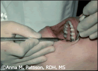
The conventional intraoral finger rest can be used effectively in most regions of the mouth with the exception of the maxillary posterior regions. Using intraoral fulcrums in these regions results in awkward positions that require wrist deviation or flexion and finger-flexing activations that split the grasp fingers from the fulcrum finger. For example, when attempting to establish an intraoral finger rest for the maxillary molars, often the middle and ring fingers must be separated in order to extend the blade back far enough to permit proper positioning of the instrument shank. This separation is necessary for proper working angulation and for insertion to the depth of the pocket (Figure 1).
The separation of the fingers is far from the ideal of a “built-up” fulcrum. It can force the clinician to activate scaling strokes with finger-flexing motion. Finger-flexing with a split fulcrum can result in less power, less accuracy, and reduced control.
Finger-flexing strokes tend to exert more lateral pressure at the beginning of the stroke and to taper off as the stroke moves in a coronal direction, making long root planing strokes with even lateral pressure difficult to achieve.
This dilemma of the maxillary posterior teeth can be solved by using extraoral hand rests that enable proper instrument placement, correct angulation, and wrist motion. Clinicians often use extraoral fulcrums for the maxillary posterior teeth with probes and explorers to enhance access to hard-to-reach surfaces, such as the distals of second or third molars. Also extraoral hand rests are routinely used during ultrasonic instrumentation. Often clinicians may not realize that the superior access attributed to ultrasonic scaling is directly related to the extraoral fulcrums being used rather than just the design of the ultrasonic tips. Some clinicians always use extraoral fulcrums for ultrasonic scaling but limit themselves to only intraoral fulcrums with hand instruments. When properly established, extraoral fulcrums can allow optimal access and angulation while providing adequate stabilization for either ultrasonic or hand instrumentation. Extraoral fulcrums differ from intraoral fulcrums because the front or back surfaces of the fingers and hand provide the support rather than the tips or pads of the fingers as with intraoral finger rests.
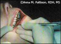
Often, the obstacle to achieving effective extraoral fulcrums results from a misunderstanding or misinterpretation of the elements that comprise a properly established extraoral fulcrum. Some clinicians believe that extraoral fulcrums should not be used because they are too unstable and may result in slipping and injury to the patient. This is true if an extraoral fulcrum with a one-point finger rest is used. A one-point extraoral finger rest established by the ring finger on the chin is not stable enough to insure adequate control. Instead, as much of the front or back surface of the fingers and hand as possible must be placed on the patient’s face to provide the greatest degree of stability. Figures 2 and 3 show two effective extraoral hand rests.
The palm-up hand rest in Figure 2 is established by resting the backs of the fingers-not the pads or tips-firmly against the skin overlying the lateral aspect of the mandible on the right side of the face. Strokes are activated by pulling in a coronal direction with the arm, not by flexing the fingers. The movement is initiated at the shoulder. The fingers remain stationary in their grasp position as the arm activates the working stroke.
The extraoral palm-up hand rest is sometimes described as a knuckle rest but this can be misleading because this technique does not actually entail curling the fingers inward to make a fist. Instead, a properly established palm-up extraoral fulcrum will position the middle, ring, and little fingers straight and together. As much of the backs of these fingers as possible, not just the knuckles, should be kept in firm contact with the basal portion of the mandible for stability and control.
The palm-down hand rest in Figure 3 is established by resting the front surfaces of the fingers flatly against the skin overlying the lateral aspect of the mandible on the left side of the face. Whenever possible, the palm of the hand should closely cup the mandible to provide additional support.
Elements of the Extraoral Fulcrum
There are three essential elements for a stable extraoral hand rest. The first element is a broad surface area of contact between the hand and the patient’s face. Maximize this surface area whenever possible to establish a secure foundation.
The second element is adequate pressure for the extraoral fulcrum. If a firm working stroke is activated against the tooth without adequate pressure exerted against the skin of the face, the stroke will lack control. It is actually the bone and soft tissue of the patient’s face that serve as the fulcrum to stabilize the working stroke. The clinician must apply sufficient pressure to the skin of the face to reach that bony foundation in order to control the instrument. Extraoral hand rests are thus fully dependent on a secure hand rest that uses pressure equal to that exerted on the tooth by the blade. The degree of pressure used varies according to the type of stroke being activated. Light-pressured assessment strokes require only light-pressured hand rests. Moderate-pressured debridement strokes call for moderate-pressure exerted onto the patient’s skin. Heavy calculus removal strokes necessitate stable, secure, firm-pressured hand rests.
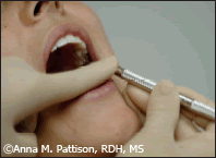
The final element of effective extraoral hand rests is an extended grasp. If the instrument is grasped too close to the working end, the resting fingers will pinch the patient’s lip against the teeth resulting in pain for the patient and instability for the clinician (Figure 4).
In a proper extraoral fulcrum, it is critical to locate the grasp on the handle away from the working end. Without the extension of the grasp, the instrument will not reach the tooth. Clinicians accustomed to intraoral finger rests are initially reluctant to use an extended grasp with an extraoral fulcrum. This stems from a common misconception that control of the instrument is dependent on a choked up grasp, close to the working end of the instrument. In fact, it is the fulcrum pressure rather than the grasp position that determines whether or not a stroke will be well controlled.
In summary, an effective extraoral fulcrum incorporates three elements:
- An extended grasp;
- A broad surface area of contact between the hand and the patient’s face; and
- Appropriate pressure onto the skin of the patient’s face prior to activation of working strokes.
Reinforced Rests
Extraoral fulcrums can also be stabilized using reinforced rests. This is when the finger or thumb of the nonoperating hand is applied to the handle or shank of the instrument during working strokes. This type of reinforcement is most commonly employed with opposite-arch finger rests and extraoral hand rests whenever precise control and pressure are compromised by the longer distance between the fulcrum and the curet blade. The reinforcing finger acts as a support that provides added control and pressure against the tooth. Direct vision must be used with reinforced rests because the nonoperating hand is not free to use the mirror. Figure 5 shows an example of a reinforced rest.
The advantages of using extraoral fulcrums far outweigh the minor technique adjustments that are required. They permit positioning that allows for precise activation with ideal angulation in areas where this is typically difficult to achieve. They have the additional advantage of using neutral wrist positions and hand-forearm pull activations and are likely to guard against damage to the muscles, tendons, and nerve sheath running through the clinician’s wrist when scaling and root planing.
As clinicians are challenged by treating more patients with loss of attachment and heavy subgingival deposits, they will no doubt begin to depend on the extraoral fulcrum as a valuable and necessary means of efficiently and reliably achieving successful outcomes. For this reason, reaching a level of competence with the extraoral fulcrum is a worthwhile endeavor for any clinician.
This article was adapted from Pattison A, Matsuda S, Pattison G. Periodontal Instrumentation . 3rd ed. Upper Saddle River, NJ: Prentice Hall. In press.
From Dimensions of Dental Hygiene. October 2004;2(10):20, 21-23.
