Concepts for Modern Implantology
Dimensionsspeaks with Paul Fletcher, DDS, about key developments in implant restorations.
Q How does peri-implantitis differ from periodontitis?
A Peri-implantitis and periodontitis present as clinically similar lesions, and research shows that bacteria present in peri-implant disease are the same as those responsible for the development of periodontitis.1 However, the progression of the diseases can differ both radiographically and histopathologically. While both teeth and dental implants have an epithelial adherence as the first line of defense against the apical progression of inflammation, the orientation of the suprabony connective tissue fibers is different.
With a natural tooth, connective tissue fibers run perpendicular to the root surface and actually insert into the cementum. Because connective tissue fibers cannot organically attach to the dental implant, the fibers tend to run to the implant, veer away from the surface, and then encircle it. Additionally, while a periodontal ligament exists between a tooth root and bone, there are no ligament fibers between bone and a titanium implant abutment—only a thin zone of glycoprotein ground substance that abuts the oxide layer of the implant.2
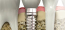
The implications of these differences are evident when examining periodontitis and peri-implantitis lesions clinically and viewing them radiographically. Because inflammation tends to follow the course of the connective tissue fibers, periodontal infrabony lesions are site specific when viewed radiographically (eg, discreet vertical lesions at any surface of a tooth). By comparison, peri-implantitis lesions tend to produce a more symmetrical, implant-specific bone loss as the breakdown follows the course of the circular fibers around the dental implant (Figure 1).
Q Are implants more susceptible to peri-implant breakdown than teeth are prone to periodontal diseases?
A Host resistance significantly impacts whether a patient will experience periodontal or peri-implant disease. Dental implants are not inherently more prone to inflammatory breakdown than natural teeth, as long as the implant surface is not exposed. If the implant surface is exposed to bacteria, implants with rough surfaces are more susceptible to peri-implant disease than implants with smooth surfaces. Implant manufacturers recognize the numerous advantages of using a rough-surface dental implant, such as speed and amount of integration, and they are actively developing implants with the ideal combination of surface roughness and resistance to periimplant disease.
Peri-implant disease research has determined that once the lesion is initiated, the inflammatory infiltrate in the peri-implantitis lesion tends to extend closer to the crest of bone than the periodontal lesion.3 Because an implant has no ligament to provide blood supply, the capillary circulation to the connective tissue fibers is not as robust as the circulation around a tooth. In the latter, the perpendicular supra-crestal connective tissue fibers and infection-fighting cells from the blood vessels in the ligament attempt to form a barrier to prevent the penetration of the lesion directly to the bone. This does not seem to occur to the same extent around a dental implant.
Q Are individuals who have had severe periodontitis more prone to periimplantitis?
A Indiividuals who show a genetic predisposition or a lack of host resistance to periodontal disease are more likely to develop peri-implantitis.
A systematic review of 15 studies where dental implants were placed in partially edentulous patients made the following observations: When patients with histories of periodontitis were compared with individuals who were periodontally healthy, the patients with histories of periodontal disease had significantly greater probing depths, more peri-implant marginal bone loss, and a higher incidence of periimplantitis.4 It was concluded that the percentage of implant survival was still acceptable if the patient was in an ongoing maintenance program.
A review of nine studies on the success of implants in patients with histories of aggressive periodontitis also concluded that bone loss occurred more often in these patients than around implants in periodontally-healthy patients or in patients with histories of chronic periodontitis.5 The studies also showed the survival rates for these implants were good if the implants were well maintained, thus they recommended that all periodontal disease be controlled prior to dental implant placement. Because the effect of questionable teeth on the long-term success of implants in patients with prior severe periodontitis is still unknown, the level of potential pathogens must be minimized to reduce the opportunity for bacteria to colonize the implants and initiate peri-implant disease.6
Q What is the latest research on the use of specific instruments for implant debridement?
A The original screw-type implant, first manufactured in Sweden during the 1980s, was composed of Type 1 commercially pure titanium. A relatively soft metal, it scratched easily when cleaned with a metal instrument harder than itself.
Today’s dental implants are made out of a harder, commercially pure Type 4 titanium, or a titanium alloy. Debridement and instrumentation are still carried out by implant-safe hand instruments, such as plastic and graphite scalers and gold- and titanium-tipped curets. Ultrasonic instruments can also be used as long as the metal tips are shielded with a plastic covering or special inserts or tips are used. No single instrument has proven superior.
The objective of therapy is to remove the plaque, calculus, endotoxins, and dead bacterial cell walls from the super osseous aspect of the implant without scratching it or the abutment surface. This is important because scratched surfaces are more susceptible to plaque and calculus accumulation.
Thoroughly removing debris from a subgingival implant surface is challenging because it can be extremely difficult to get 360° around an implant that is often splinted and covered by a superstructure. An ultrasonic instrument with a safe tip is often useful in these situations.
QWhat strategies are helpful in treating ailing implants?
A Peri-implant disease can be separated into two entities: mucositis and peri-implantitis. Mucositis is comparable to gingivitis around teeth. Bleeding is present on probing, and pocket depths can be 5 mm or greater as long as there is no evidence of increasing pocket depth or progressive radiographic bone loss beyond the first year of placement. Treatment is directed toward controlling inflammation and decreasing the bacterial load. Meticulous patient oral hygiene along with antimicrobial mouthrinses, mechanical debridement, subgingival irrigation, and local or systemic drug delivery are beneficial in reducing mucositis and preventing its progression to periimplantitis.7
When pocketing continues to increase and progressive bone loss is visible radiographically, the implant is considered to have peri-implantitis. Research shows that peri-implantitis cannot be predictably treated nonsurgically. Open flap debridement is needed to gain access to the implant to allow for surface detoxification. Implant detoxification can arrest the disease process by reducing inflammation and creating a surface that will allow for re-adherence of the gingiva to the implant. Presurgical systemic antibiotics, followed by chlorhexidine application, surface burnishing with saline, and laser therapy are all used for implant disinfection. Bone grafting of the osseous defect, with and without membranes, has also been attempted. While studies show bone filling into the outer aspects of the defect, the amount of bone that reintegrates to the implant is limited.8 When treating peri-implant disease, any benefit obtained can be lost without a continuous maintenance program and impeccable oral hygiene.
Q What does the future hold for the treatment of peri-implantitis?
A Dentistry is beginning to acknowledge that the prevalence of periimplant disease is greater than originally thought. Early interceptive therapy is becoming more common among patients who have lost their teeth from periodontal disease; the sooner the condition is recognized, the more successful the treatment.7 Baseline probing measurements should be taken 3 months after placement of the final restoration, and at all subsequent recall appointments. As probing depths are often 5 mm or greater initially in areas with thick gingiva, an increase in pocketing along with bleeding on brushing or probing are warning signs that should not be ignored. For individuals who have an aggressive response to plaque, 5-mm pocketing alone should be an indication to initiate treatment. Radiographs should be taken at the insertion of the restoration— and at 6-month intervals if evidence of progressive peri-implant disease is present. Currently, researchers are looking at multiple new approaches, including electrochemical disinfection, to detoxify an implant surface. The predictable and successful treatment of periimplant disease is still in the development stage.
Q What’s on the horizon for implant design?
A Each implant manufacturer is racing to be the first to develop a dental implant that will integrate quickly and strongly, while also holding the marginal bone and gingiva coronally to minimize recession and optimize esthetics. To reach that end, the latest generation of implants is designed with a taper so the implants will wedge into the osteotomy site and gain initial stability from the coronal cortical bone, while also allowing for the immediate placement of a provisional restoration. Additionally, the thread design is deeper and sharper, with self-cutting edges to lock more effectively into the softer cancellous bone. While the first screw-shaped titanium implants had relatively smooth surfaces, researchers quickly realized that roughening the dental implant surface would increase the surface area available for integration. Additive techniques, such as plasma spraying with small particles of titanium and coating the implant with hydroxyapatite, were once popular. However, 10-year longitudinal studies showed these implants were more susceptible to peri-implant disease than smooth-surfaced implants, and they were eventually taken off the market. Manufacturers roughen today’s implants using subtractive techniques such as acid etching or sand blasting, or a combination of the two. The surfaces have microscopic notches and grooves in them. Research has shown that some coronal thread designs have actually had connective tissue fibers mechanically locked into them. While these implants are more osteoconductive and integrate faster than smooth-surfaced implants, time will tell if they are as resistant to peri-implant disease.
Other future trends include implants made out of nonmetallic materials such as ceramics, carbon, and composites. Additionally, implants will be multi-surfaced, where a different surface may be used for the top, middle, and bottom. Thus, in a patient with soft bone and a history of periodontitis, the most coronal part of the implant may have a machined surface resistant to peri-implantitis, the middle third may have a moderately rough sand-blasted surface, and the bottom third may have an hydroxyapatite coating, which is the only surface that biologically bonds to bone.
REFERENCES
- Mombelli A. Microbology and antimicrobial therapy of peri-implantitis. Periodontol 2000. 2002;28:177-189.
- Lindhe J, Berglundh T, Ericsson I, et al. Experimental breakdown of peri-implant and periodontal tissues. A study in the beagle dog. Clin Oral Implants Res. 1992;3:9-16.
- Berglundh T, Lindhe J, Ericsson I, et al. The soft tissue barrier at implants and teeth. Clin Oral Implants Res. 1991;2:81-90.
- Karoussis IK, Kotsovilis S, Fourmousis I. A comprehensive and critical review of dental implant prognosis in periodontally compromised partially edentulous patients. Clin Oral Implants Res. 2007;18:669-679.
- Quirynen M, De Soete M, van Steenberghe D. Infectious risks of oral implants: a review of the literature. Clin Oral Implants Res. 2002;13:1-19.
- Greenstein G, Lamster I. Bacterial transmission in periodontal diseases: a critical review. J Periodontol. 1997;68:421-431.
- Zitzmann NU, Berglundh T. Definition and prevalence of peri-implant diseases. J Clin Periodontol. 2008;35(Suppl 8):286-291.
- Nibali L, Donos N. Radiographic bone fill of peri-implantitis defects following nonsurgical therapy: report of three cases. Quintessence Int. 2011;42:393-397.
From Dimensions of Dental Hygiene. September 2011; 9(9): 44, 46-48.
