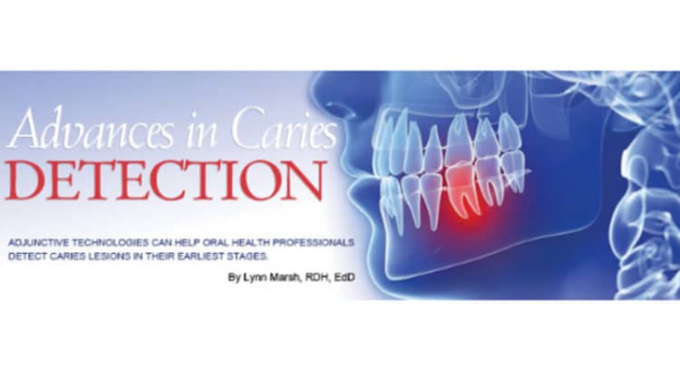
Advances in Caries Detection
Adjunctive technologies can help oral health professionals detect caries lesions in their earliest stages.
Dental caries is one of the most prevalent early childhood diseases, and its prevention remains challenging. Strategies for reversing caries lesions in their initial stages are available, but early detection is key to this treatment approach. New technologies that detect initial stages of enamel demineralization are now available. The purpose of early caries diagnostic methods is to offer objective information about the presence and severity of caries lesions, complement oral health professionals’ subjective assessment, and provide evidence-based clinical caries diagnosis.1 Supplementing traditional caries diagnostic methods with sophisticated, more perceptive tools may help improve patient outcomes.
LIGHT-INDUCED FLUORESCENCE
Current diagnostic methods can detect caries but they are unable to quantify the mineral condition of a lesion. Light-induced fluorescence measures changes seen in the demineralized enamel compared to surrounding sound enamel, using these measurements to determine the amount of mineral lost during demineralization.2 The images—created utilizing tooth fluorescence—help ascertain the location and depth of caries lesions.
In addition to early caries diagnosis, light-induced fluorescence can also be used to evaluate sealants for sound margins. While sealants are placed on occlusal surfaces to prevent the development of caries lesions in the pits and fissures of posterior teeth, caries can form under a visually intact sealant. This distinction between a sound sealant and a compromised sealant is almost impossible to make using the traditional methods of tactile and visual examinations. An existing sealant will fluoresce, however, when light-induced fluorescence is used, indicating the sealant is compromised.
INFRARED FLUORESCENCE
With the increased use of fluoride mouthrinses and toothpastes and professional application of fluoride varnish, the pattern of caries has also changed. Pit and fissure caries are now the most common type of decay.3 Although fluoride helps reduce enamel demineralization, caries lesions that advance into the dentin progress underneath a clinically intact enamel surface. Given the difficulty of visually inspecting the pits and fissures of molars, cases of occlusal dentinal caries are commonly missed on visual examination, leading to the diagnosis of caries lesions during the late stage of disease.3
Infrared fluorescence is a noninvasive, pain-free technique used to detect early occlusal caries lesions otherwise undiagnosable through traditional methods. This modality emits an infrared light that can be absorbed by organic and inorganic tooth materials, and the process of remitted fluorescence shows various scales between 0 and 99. Infrared fluorescence measures the amount of laser fluorescence within the tooth. Healthy teeth will have little to no fluorescence. But if a tooth contains caries, more laser light is fluoresced. Compared to baseline readings, a higher reading indicates the presence of a caries lesion within the structure of the tooth. The amount of laser light reflected back correlates with the amount of decay within the tooth. A value of 20 to 25 or higher designates a caries lesion. The higher the fluorescence value, the deeper the lesion.4
INFRARED FLUORESCENCE COMBINED WITH PHOTOTHERMAL RADIOMETRY
Photothermal radiometry (PTR) is used to detect mechanical holes and demineralized enamel in interproximal contact areas.5 The interproximal area of a tooth is a common region for demineralization, due to dental biofilm buildup. PTR is sensitive to assorted defects—such as a deep caries lesions, demineralized areas, edges or cracks, and surface stains—while infrared fluorescence exhibits low sensitivity and spatial resolution.6 This technology scans over the surface of a fissure into demineralized enamel and dentin, which shows higher amplitude than healthy teeth.
Garcia et al7 found a positive correlation between PTR results and mineral loss or lesion depth measured with transverse microradiography. These results indicated that PTR is capable of monitoring artificially created caries lesions, their revolution during demineralization, and the reversal of lesions during remineralization.7 The use of PTR as an adjunctive tool for early caries diagnosis demonstrates consistent variations in the presence of demineralized interproximal tooth structures.
TRANSILLUMINATION
Conventionally, oral professionals rely on visual and tactile examinations, in addition to dental radiography, for caries detection. Consequently, incipient caries lesions in interproximal areas may be missed. In the search for more accurate diagnostic approaches, investigators have developed noninvasive methods for early caries detection based on dental tissue optics.
Transillumination of dental enamel with near-infrared light is a promising optical imaging technique for detecting the presence of early dental caries lesions and measuring their severity.8 The visual properties of a caries lesion are distinctive from those of the surrounding healthy tooth surfaces. Transillumination intensifies the change in scattering and absorption of light photons in the caries lesion, thereby making it appear as a dark shadow.9 Another benefit is that transillumination can be used for caries detection on all tooth surfaces with sensitivity in the interproximal areas.
ALTERNATING CURRENT IMPEDANCE SPECTROSCOPY
Alternating current impedance spectroscopy involves the measurement of electrical resistance of the tooth structure. This technology applies a small alternating electrical signal through the tooth and monitors the response. By changing the frequency of the applied signal, a spectrum is captured that provides valuable insight into the physical and chemical properties of the tooth.10 Alternating current impedance spectroscopy facilitates a more precise detection of early caries lesions not clinically visible during an intraoral examination.
PRACTICAL APPLICATION
Accurate assessment of the presence or absence of disease is essential not only in dentistry, but all health care environments. Visual, tactile, and radiographic examinations are the traditional means to diagnosing dental caries. The aforementioned caries detection technologies can improve the sensitivity of early caries diagnosis, and they may support the detection of questionable surfaces, but they are not fool-proof. A review of the evidence supporting these caries detection technologies is decidedly mixed.11–14 Some are likely to produce false positives, while others may miss lesions occurring around restorations. While these early caries diagnostic methods may be valuable adjunctive technologies, they should be used in addition to traditional diagnostic methods.
REFERENCES
- Karlsson L. Caries detection methods based on changes in optical properties between healthy and carious tissue. Int J Dent. 2010;2010:270729.
- Amaechi BT, Higham SM. Quantitative light-induced fluorescence: a potential tool for general dental assessment. J Biomed Opt. 2002;7:7–13.
- Chu CH, Lo EC, You DS. Clinical diagnosis of fissure caries with conventional and laser induced fluorescence. Lasers Med Sci. 2010;25:355–362.
- Bahrololoomi Z, Musavi SA, Kabudan M. In vitro evaluation of the efficacy of laser fluorescence (DIAGNOdent) to detect demineralization and remineralization of smooth enamel lesions. J Conserv Dent. 2013;16:362–366.
- Jeon RJ1, Hellen A, Matvienko A, Mandelis A, Abrams SH, Amaechi BT. In vitro detection and quantification of enamel and root caries using infrared photothermal radiometry and modulated luminescence. J Biomed Opt. 2008;13:034025
- Jeon RJ, Matvienko A, Mandelis A, Abrams SH, Amaechi BT, Kulkarni G. Detection of interproximal demineralized lesions on human teeth in vitro using frequency-domain infrared photothermal radiometry and modulated luminescence. J Biomed Opt. 2007;12:034028.
- Garcia J, Mandelis A, Abrams SH, Mativenko A. Photothermal radiometry and modulated luminescence: applications for dental caries detection. In: Handbook of Biophotonics, Vol 2: Photonics for Health Care. Popp J, Tuchin VV, Chiou A, Heinemann SH, eds. Hoboken, NJ: Wiley; 2011:1047.
- Maia AM, Karlsson L, Margulis W, Gomes AS. Evaluation of two imaging techniques: near-infrared transillumination and dental radiography for the detection of early approximal enamel caries. Dentomaxillofac Radiol. 2011;40:429–433.
- Astvaldsdóttir A, Ahlund K, Holbrook WP, de Verdier B, Tranæus S. Approximal caries detection by DIFOTI: in vitro comparison of diagnostic accuracy/efficacy with film and digital radiography. Int J Dent. 2012;2012:326401.
- Ari T, Ari N. The performance of ICDAS-II using low-powered magnification with light-emitting diode headlight and alternating current impedance spectroscopy device for detection of occlusal caries on primary molars. ISRN Dent. 2013;14:276070.
- Braga MM, Ekstrand KR, Martignon S, Imparato JC, Ricketts DN, Mendes FM. Clinical performance of two visual scoring systems in detecting and assessing activity status of occlusal caries in primary teeth. Caries Res. 2010;44:300–308.
- Ferreira Zandona A, Zero DT. Diagnostic tools for early caries detection. J Am Dent Assoc. 2006;137:1675–1684.
- Pretty IA. Caries detection and diagnosis: novel technologies. J Dent. 2006;34:727–739.
- Berg J. Addressing the silent epidemic. Dimensions of Dental Hygiene. 2012; 10(9):42–45.
From Dimensions of Dental Hygiene. May 2014;12(5):42,44.
