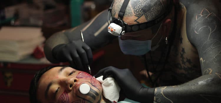
Extreme Body Modifications
Providing effective care and recommendations to this patient population is key to supporting oral health.
This course was published in the March 2018 issue and expires March 2021. The authors have no commercial conflicts of interest to disclose. This 2 credit hour self-study activity is electronically mediated.
EDUCATIONAL OBJECTIVES
After reading this course, the participant should be able to:
- Discuss the prevalence of body modifications in the United States, in addition to their historical and cultural significance.
- Identify the types of extreme body modifications and their risks.
- Explain the role of oral health professionals in treating this population.
Body modifications, such as tattoos, piercings, and branding/scarification, have been used by humans for thousands of years. While acceptance of these practices varied in different cultures, their social acceptance has been growing globally and in the United States.1 In 2016, approximately one in three Americans had a tattoo—a 20% increase from 2012.2Nearly four in 10 Millennials had at least one tattoo and one in four had a piercing in a place other than earlobes, according to a 2010 Pew Research Center report.3 The majority of Americans feel comfortable with seeing individuals with tattoos in various professional roles.2 A number of more extreme trends, such as skin scarification, sclera tattooing, and tongue bifurcation, have emerged. Few studies have been conducted on the effects of these modifications; however, media reports, case studies, and personal accounts offer evidence of complications. Oral health professionals should be familiar with these body modifications and understand their possible local and general complications, implications for patient assessment and treatment, and strategies to minimize risks.
HISTORY AND CULTURAL SIGNIFICANCE
Body modifications are associated with cultural expression in religious, social, punitive, sexual, esthetic, and medical contexts.1,4,5 One of the oldest human specimens with tattoos was dated at more than 5,200 years old. The markings were found on the lower spine, wrists, knee, and ankle joints, and it is speculated that they were made for therapeutic purposes.5–7 Egyptians used tattoos to signify social or religious status, and tattoos on the abdomen and thighs were used to ward off evil during birthing.7 Slaves and prisoners were known to be permanently marked in early Greek and Roman societies and South Pacific cultures have long used ornamental tattoos.6
Body piercing of the lip, ears, and tongue trace back 4,000 years to 5,000 years and were popular aesthetic and tribal adornments in ancient Africa. Nobility in ancient Egypt pierced their navels, while Roman soldiers pierced their nipples to demonstrate virility.8 Christians in the Middle Ages feared the plague and discouraged the practice. The art of the Renaissance and traveling Europeans revived the practice, which waned somewhat in the 1920s in the US, then came back with a vengeance in the 1960s and 1970s, and has become mainstream in current times.5
Scarification, the purposeful manipulation of healing to create markings on the skin, has ancient roots in some cultures. It is believed to have originated in the Australian Aborigines as early as 60,000 BC.5 While currently prevalent in some communities as a method of cultural self-expression and identification, as well as for medicinal purposes by placing herbs into the cuts for the prevention of various diseases and snake bites,5 there are no studies on the rates of this practice in the US.1 This more extreme body art practice emerged in San Francisco in the 1980s and increased in popularity in the 1990s.5,9
LAWS AND REGULATIONS
The Association of Professional Piercers (APP) states its support for “the right for all adults to adorn or modify their bodies in a safe, informed, and consensual manner when performed by a qualified practitioner under appropriate asepsis.”10 APP and the Alliance of Professional Tattooists (APT)11 offer detailed information on safe practices, dangers, and complications and their prevention, as well as links to the state legislatures that regulate various body art forms. In the absence of federal regulations, all states except Nevada have adopted laws addressing body art to ensure safety, protection of minors, and to prevent conflict with the scope of practice of medical and dental professionals.12 However, most regulations address body art limited to tattooing and piercings, with several states including branding/scarification, sub-/transdermal implants, and tongue bifurcation.12 Scarification is not addressed in 19 states, while other states’ legislation ranges from prohibition of the practice to some degree of regulation.1 Currently, Oklahoma is the only state that bans sclera tattooing and the Indiana legislature is considering it.13
TYPES OF MODIFICATIONS
Sclera tattooing. Eyeball tattooing, specifically corneal tattooing, is an ancient practice that has specific medical indications for patients with congenital eye defects, such as aniridia (absence of the iris), cataracts, and scars due to traumatic injury to the eye.14 This complex surgical procedure can be safely performed by trained ophthalmologists and can result in improved appearance, although ocular function is not restored.
On the contrary, cosmetic sclera tattooing is performed by self-taught tattoo artists without anesthesia in a nonmedical environment.15 Luna Cobra, the inventor of cosmetic sclera tattooing who first tried the procedure in 2007, claims to have since refined the technique. Even though he reports no complications, Cobra admits there are risks.16 The American Academy of Ophthalmology (AAO) emphasizes that cosmetic sclera tattooing has not been studied and strongly advises against it. The AAO warns of serious risks, including infection from injection or ink, decreased vision, complete blindness, and potential loss of the eye.17 A video case report of a 24-year-old patient demonstrates that despite surgical intervention and removal of some of the black tattoo ink, followed by antibiotic treatment, the patient’s eye was subsequently removed due to intractable pain.18
http://www.ajocasereports.com/article/S2451-9936(16)30143-8/fulltext
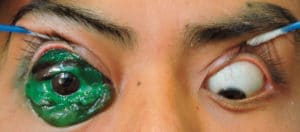
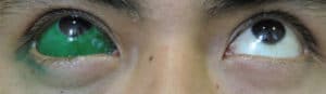
Published cases describe individuals seeking professional medical care several days19,20 to several weeks15 after sclera tattooing. Their chief complaints ranged from conjunctival lumps/nodules without reported pain15,19 to pain, photophobia, decreased vision, and eyelid edema (Figure 1).19 Different mixtures of tattoo dyes19 resulted in various coloration of patients’ scleras. In these cases, following appropriate treatment including corticosteroids and antibiotics, the patients’ condition improved (Figure 2);19 however, long-term complications, such as secondary glaucoma20 and conjunctival granulomas that can lead to scleral thinning or malignancy, remain possible.15,19 Risks during the procedure performed by untrained personnel and without the use of a surgical microscope can result in globe penetration,15,20 traumatic cataract, retinal detachment, and endophtalmatis.15 As the tattoo ink acts as a foreign body, it can cause ocular inflammatory and allergic reactions to the metallic elements, such as nickel, cobalt, copper, chromium, and iron.19 Until recently, sclera tattooing has been relatively unknown outside of a small group of practitioners and their clients, and the medical professionals who treat the resulting complications.15,17–20
http://www.ajocasereports.com/article/S2451-9936(16)30143-8/fulltext
Scarification. This is the practice of creating permanent scars by cutting, burning, or branding images, words, or patterns in the skin.1,5 It can be performed by cutting with scalpel or knife, hot or cold branding (using liquid nitrogen), thermo- and electro-cautery, and moxibustion (placing incense on the skin and allowing it to burn until it is extinguished in the underlying layers).1,5 Strips of skin can be removed to produce larger patterns, and wounds can be irritated by chemicals (iodine, citrus juice) and scraping off scabs to enhance scar formation.1,21 Regardless of the method, results can be inconsistent due to unpredictable healing and possible complications such as infections and keloid formation.1 Performed without anesthesia and with intentionally prolonged healing time, this procedure requires remarkable pain tolerance.
Scarification and other more dramatic, intense, and numerous body modifications can be a sign of a nonsuicidal self-injury syndrome (NSSI), a disorder associated with mental health conditions such as psychotic, personality, and anxiety disorders. The prevalence of NSSI in adolescents is estimated at 14% to 24%.1 Careful, nonjudgmental interviewing by a health care professional observing these body modifications can help determine the intent. Clinicians need to distinguish between normal body modifications and self-injurious behaviors that can be a symptom of underlying mental health disorders, and help identify individuals at risk for greater self-harm, including suicide.1,4,21 Because many body modifications, including scarification, involve the head/neck area and face, oral health professionals may play an important role.
GWEN COHEN BROWN, DDS, FAAOMP, AND COURTESY OF THE DENTAL HYGIENE DEPARTMENT OF THE NEW YORK CITY COLLEGE OF TECHNOLOGY/CUNY
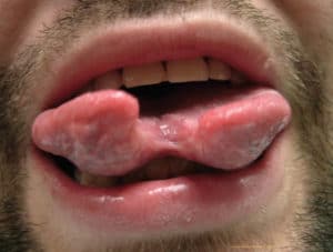
Tongue bifurcation. Also known as tongue splitting, forking, or bifid tongue, this modification involves separating the tongue anteriorly into two parts of various length, to resemble a snake’s tongue (Figure 3). This procedure can be done by cutting with a scalpel or laser beam along the midline and cauterizing or suturing the edges to prevent reattachment.21,22 Tongue bifurcation has also been reported as an unwanted complication of an infected tongue piercing that successfully healed after a surgical intervention.23 Tongue splitting is an extremely painful procedure, which is typically done by medically untrained artists with inadequate or no anesthesia.21,22,24 It can result in infection, inflammation, blood loss due to high vascularization of the tongue, and nerve damage that can persist even in well-healed bifid tongue.22,24 With training, individuals can learn to control the two parts of their tongue independently and the effects on speech may be unnoticeable.21,22 The American Dental Association (ADA) advises against tongue bifurcation as an invasive and dangerous procedure performed for nonmedical reasons.25
Tongue piercings can precede or accompany tongue bifurcation, and they can have serious complications and implications for oral health care. More frequent in women, tongue piercing is the most prevalent oral/perioral piercing (5.6%).8 It has been associated with rare but serious complications including cerebellar abscess;26 infective endocarditis;27 local infections; and increased incidence of tooth injuries, such as enamel fissures, fractures, and gingival recession, especially in the mandibular lingual incisor area.28Unfortunately, surveys of piercers29 and clients with oral piercings30,31 showed low awareness of the associated local and systemic risks, including possibility of transmission of blood-borne diseases, bacterial infections, temporary and permanent paralysis due to possible nerve damage, and local complications. Piercers also demonstrated poor knowledge of oral cavity and tongue anatomy,29and post-procedural care recommendations were limited to using mouthrinse, while follow-up visits with piercing specialists or medical professionals were recommended very rarely.30,31
CONSIDERATIONS FOR ORAL HEALTH PROFESSIONALS
Assessment and documentation of the status of orofacial modifications should be performed as part of the comprehensive intra- and extraoral examination and include the location, description, and condition of sites and surrounding hard and soft tissues. Photographic documentation is advisable for future comparison. Familiarity of the oral health professional with typical post-procedural healing time and potential adverse reactions ensure safety and create an environment of respect and candor in the dental setting.4
The routine removal of all metal objects and jewelry in the field of view of a diagnostic image is essential. In panoramic imaging, a metal object obscures anatomy and potential pathology in its own location, and creates an enlarged ghost image on the contralateral side. The complexity of cone-beam computerized tomography imaging causes artifacts when imaging dense metals obscuring anatomical structures adjacent to the metal object, and create multiple light/dark bands across the image layers. Many software programs can reduce these effects from dental materials in the dentoalveolar area, but may be less able to eliminate these effects in perioral and facial piercings.32,33 Recently placed, implanted or soldered objects may preclude or require planned removal by trained personnel with vulnerable areas maintained with sterile tubing or sutures.4
In magnetic resonance imaging (MRI), minor burns have been associated with tattoos,7 although a recent study with a new MRI machine showed safety and effectiveness with no side effects associated with tattooed regions.34 Artifacts on MRIs, however, have been discovered from even small tattoos, such as permanent eye liner.7
During dental procedures, facial, perioral, and mucosal piercings need not be removed unless they directly interfere with the treatment site, affect asepsis, or limit the use of isolation or suction devices. Removed hardware may be cleaned and disinfected as needed, and vulnerable openings maintained with nonmetallic instruments.4,35,36 During medical procedures involving intubation, intraoral jewelry may pose a significant risk of injury or aspiration.
Oral health professionals should assess patient awareness of hard and soft tissue risks and proper care of tongue and lip piercings, with the tongue being the most common, and problematic.28,30 Healing time for tongue piercing and bifurcation procedures is estimated at 4 weeks to 6 weeks and lips at 6 weeks to 8 weeks.4 Common acute complications of tongue piercing include hypersensitive reactions to metal, swelling, and pain, and, less commonly, increased salivary flow, severe infection, airway obstruction, severe bleeding, embedding of the object, and nerve damage. Trauma to teeth includes chips, fractures, pulpal damage, and gingival recession. Localized hyperplastic growth is not uncommon.25,30
The ADA and the American Academy of Pediatric Dentistry strongly discourage intraoral piercings, but recognize the oral health professional’s role in assessment, care, and education.25,37 Patients should be informed about maintaining excellent oral hygiene practices after piercing procedures and recognizing abnormal swelling, prolonged bleeding, pain, and exudate.25
CONCLUSION
Discussion regarding the care and complications associated with body modifications that may affect health or treatment maximizes safety and effectiveness. Oral health professionals with knowledge and recommendations about modifications are able to build trust. For young patients, anticipatory guidance may prevent future issues and even influence decisions, which have been shown to be often made in haste and without adult input. Discussion may focus on the motivation and permanency of the procedure, associated risks, difficulty of removal, need for asepsis, and choice of a qualified professional to perform the procedure.38 Especially dangerous procedures, such as sclera tattooing and tongue bifurcation, should be discouraged. For adolescents and adults, information on the care and complications associated with various body modifications delivered objectively from a knowledgeable provider may be received with openness.
All forms of body modification are growing in prevalence and social acceptability, assuring that oral health professionals will encounter patients exhibiting these phenomena and their associated risks and complications. Preparedness in current knowledge and communication skills will assist clinicians in becoming a reliable resource for these patients.
REFERENCES
- Breuner CC, Levine DA; Committee On Adolescence. Adolescent and young adult tattooing, piercing, and scarification. Pediatrics. 2017;140:e20163494.
- The Harris Poll. Tattoo Takeover: Three in Ten Americans Have Tattoos, and Most Don’t Stop at Just One. Available at: theharrispoll.com/health-and-life/Tattoo_Takeover.html. Accessed February 14, 2018.
- Pew Research Center. Millennials: A Portrait of Generation Next Confident. Connected. Open to Change. Available at: pewsocialtrends.org/2010/02/24/millennials-confident-connected-open-to-change/ Accessed February 14, 2018.
- Dunn D. Body art and the perioperative process. AORN J. 2016;104:326–340.
- Perper M, Aldahan AS, Tsatalis JP, Nouri K. Modifications of body surface: piercings, tattoos, and scarification. Int J Dermatol. 2017;56:351–353.
- Lineberry C. Tattoos: the ancient and mysterious history. Available at: smithsonianmag.com/history/tattoos-144038580/. Accessed February 14, 2018.
- Durkin SE. Tattoos, body piercing, and healthcare concerns. J Radiol Nurs. 2012;31:20–25.
- Hennequin-Hoenderdos N, Slot D, Van der Weijden G. The prevalence of oral and peri-oral piercings in young adults: a systematic review. Int J Dent Hyg. 2012;10:223–228.
- Guynup, S. Scarification: ancient body art leaving new marks. Available at: news.nationalgeographic. com/news/2004/07/0728_040728_tvtabooscars.html. Accessed February 14, 2018.
- Association of Professional Piercers. Safe Piercing. Available at: safepiercing.org/safe_piercing.php#mods. Accessed February 14, 2018.
- Alliance of Professional Tattooists. About Us. Available at: safe-tattoos.com/about.html. Accessed February 14, 2018.
- National Conference of State Legislatures. Tattooing and Body Piercing. State Laws, Statutes and Regulations. Available at: ncsl.org/ research/health/tattooing-and-body-piercing.aspx. Accessed February 14, 2018.
- Rudavsky S. Eyeball tattoos? Here’s why Indiana lawmaker wants to ban them. Indianapolis Star. January 8, 2018.
- Pitz S, Jahn R, Frisch L, Duis A, Pfeiffer N. Corneal tattooing: an alternative treatment for disfiguring corneal scars. Br J Ophthalmol. 2002;86:397–399.
- Brodie J, El Galhud H, Bates A. A case of episcleral tattooing—an emerging body modification trend. BMC Ophthalmol. 2015;8:15.
- Cobra L. Eyeball tattooing. Available at: http://lunacobra.net/services/eyeball-tattooing. Accessed February 14, 2018.
- Gudgel D. Eyeball tattoos are even worse than they sound. Available at: aao.org/eye-health/news/eyeball-tattoos-are-even-worse-than-they-sound. Accessed February 14, 2018.
- Freund PR, Greve M. Scleral tattoo gone wrong. Available at: aao.org/clinical-video/scleral-tattoo-gone-wrong. Accessed February 14, 2018.
- Duarte G, Cheja R, Pachón D, Ramírez C, Arellanes L. Case series: Two cases of eyeball tattoos with short-term complications. Am J Ophthalmology Case Reports. 2017;5:26–28.
- Cruz NF, Santos KS, Farah ML, Felberg S. Conjunctival tattoo with inadvertent globe penetration and associated complications. Cornea. 2017;36:625–627.
- Benecke M. First report of nonpsychotic self-cannibalism (autophagy), tongue splitting, and scar patterns (scarification) as an extreme form of cultural body modification in a Western civilization. Am J Forensic Med Pathol. 1999;20:281.
- Bressmann T. Speech adaptation to a self-inflicted cosmetic tongue split: perceptual and ultrasonographic analysis. Clin Linguist Phon. 2006;20:205–210.
- Fleming PS, Flood TR. Bifid tongue—a complication of tongue piercing. Br Dent J. 2005;198:265.
- Aga F, Harris R. Cosmetic tongue split. Br Dent J. 2013;214:275.
- American Dental Association. Intraoral/Perioral Piercing and Tongue Splitting. Available at: ada.org/en/member-center/oral-health-topics/oral-piercing. Accessed February 14, 2018.
- Martinello RA, Cooney EL. Cerebellar brain abscess associated with tongue piercing. Clin Infect Dis. 2003;36:e32–e34.
- Tronel H, Chaudemanche H, Pechier N, Doutrelant L, Hoen B. Endocarditis due to Neisseria mucosa after tongue piercing. Clin Microbiol Infect Off Publ Eur Soc Clin Microbiol Infect Dis. 2001;7:275–276.
- Hennequin-Hoenderdos N, Slot D, Van der Weijden G. The incidence of complications associated with lip and/or tongue piercings: a systematic review. Int J Dent Hyg. 2016;14:62–73.
- Vozza I, Fusco F, Bove E, Ripari F, Corridore D, Ottolenghi L. Awareness of risks related to oral piercing in Italian piercers. Pilot study in Lazio Region. Ann Stomatol (Roma). 2015;5:128–130.
- De Moor RJG, De Witte AMJC, Delmé KIM, De Bruyne MA, Hommez GMG, Goyvaerts D. Dental and oral complications of lip and tongue piercings. Br Dent J. 2005;199:506–509.
- Vozza I, Fusco F, Corridore D, Ottolenghi L. Awareness of complications and maintenance mode of oral piercing in a group of adolescents and young Italian adults with intraoral piercing. Med Oral Patol Oral Cirugia Bucal. 2015;20:e413–418.
- Liang H, Flint D, Benson B. Why should we insist patients remove all jewellery? Dentomaxillofacial Radiol. 2011;40:328–330.
- Schulze R, Heil U, Gross D, Bruellmann DD, et al. Artefacts in CBCT: a review. Dentomaxillofacial Radiol. 2011;40:265–273.
- Noureddine Y, Bitz AK, Ladd ME, et al. Experience with magnetic resonance imaging of human subjects with passive implants and tattoos at 7 T: a retrospective study. Magma N Y N. 2015;28:577–590.
- Smith FD. Caring for surgical patients with piercings. AORN J. 2016;103:583–596.
- Maspero C, Farronato G, Giannini L, Kairyte L, Pisani L, Galbiati G. The complication of oral piercing and the role of dentist in their prevention: a literature review. Stomatologija. 2014;16:118–124.
- Policy on intraoral/perioral piercing and oral jewelry/accessories. Pediatr Dent. 2017;39(6):83-84.
- Montgomery DF, Parks D. Tattoos: Counseling the adolescent. J Pediatr Health Care. 2001;15:14–19.
From Dimensions of Dental Hygiene. March 2018;16(3):38-41.
