Practical Benefits of the Intraoral Scanner
Developments in digital dentistry have resulted in an increased and improved application of three-dimensional (3D) technologies in patient assessment and treatment.
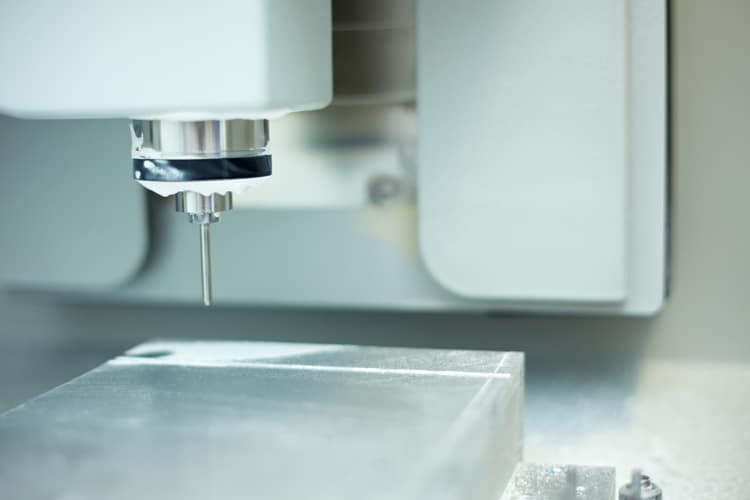
Developments in digital dentistry have resulted in an increased and improved application of three-dimensional (3D) technologies in patient assessment and treatment. For the past three decades, CAD/computer-aided manufacturing (CAM), or intraoral scanning devices, have been used in dentistry. Intraoral cameras and 3D scanners can enhance the oral health care experience. Scanners have a variety of uses, including designing precision full-ceramic crowns, veneers, inlays, and occlusal guards, as well as assisting with implants. By 1985, the first 3D intraoral imaging system became commercially available and was typically used by laboratories to fabricate precise restorations.
Photo Credit: style-photographs / iStock / Getty Images Plus
Three-Dimensional Models
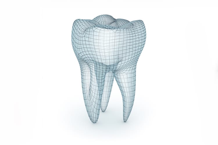
Comparable to other 3D scanners, intraoral scanners project a light source laser or structured light to scan objects, such as full dental arches, prepared teeth, and implant scanbodies. Intraoral scanners capture images of the hard and soft tissues, as well as restorations. Intraoral images are captured by the scanner and processed by software, which, in turn, generates point clouds. The point clouds are a large collection of points that generate a representation of the existing structures. They are then triangulated by the same software, producing a 3D model of the scanned range. The 3D models of the dentition and tissues are the outcome of the digital impression; they provide an alternative to traditional impression materials and stone models.
Photo Credit: alex-mit / iStock / Getty Images Plus

Full-Mouth Scans
Clinicians with appropriate knowledge and training can complete a full-mouth scan in 3 minutes to 5 minutes. Patients are immediately shown images of fractured teeth, deteriorating restorations, calculus, and gingival inflammation. Oral health professionals can monitor orthodontic relapse, malocclusion, attrition, abfraction, and any changes in gingival recession. Scans can effectively show occlusion and its relevance to oral health. Patients can see areas of heavy occlusal wear and periodontal conditions that may be related to occlusal trauma. Images can also be used to evaluate crowding, overjet, and overbite. Discussing orthodontic treatments to achieve ideal occlusion and prevent trauma is made easier by the use of intraoral scans.
Photo Credit: ayo888 / iStock / Getty Images Plus
Digital Impressions
Digital impressions decrease patient discomfort considerably when compared with conventional physical impressions. The digital scan eliminates the need for materials and impression trays, which are often uncomfortable for patients. According to the literature, patients prefer digital impressions over conventional impressions. Conventional physical impressions may cause temporary discomfort, as the technique requires the material to be in the mouth until it sets. Patients with strong gag reflexes or excess saliva and children are the least likely to tolerate conventional impression procedures. With digital impressions, If the patient is experiencing discomfort, the scan can be paused and restarted as often as needed.
Photo Credit: Feverpitched / iStock / Getty Images Plus

Training Required
Using the intraoral scanner requires training for all team members. Clinicians with a greater affinity for technology tend to adapt to the new tool more quickly. To better understand the technology and its advantages, offices utilizing intraoral scanners will need to cross-train the dental team. All dental team members need to be comfortable discussing the advantages of the scans used in the office. Knowledgeable clinicians, for example, can describe how CAD/CAM restorations can help retain maximum tooth structure and why this is important to the strength of the tooth and highlight their benefits such as one-visit restorations.
Photo Credit: asiseeit / E+

Cost Considerations
The initial cost of incorporating intraoral scanning units can be significant. When integrating a digital system into a dental practice, several factors influence cost, including the ability to mill restorations in office. Intraoral scanners can be purchased in open-format, which allows the clinician to immediately open the file once scanned. This format is preferred not only because it is versatile, but because it also reduces future costs incurred in a closed format, such as the need to purchase specific licenses and pay an annual or monthly fee to unlock files. Clinicians who lack computer and software integration skills might opt to purchase a closed format. Closed systems are fully proprietary and do not integrate with other components from other companies. Understanding the different technologies available, along with the expectations of the clinician, are important when selecting the proper device.
