

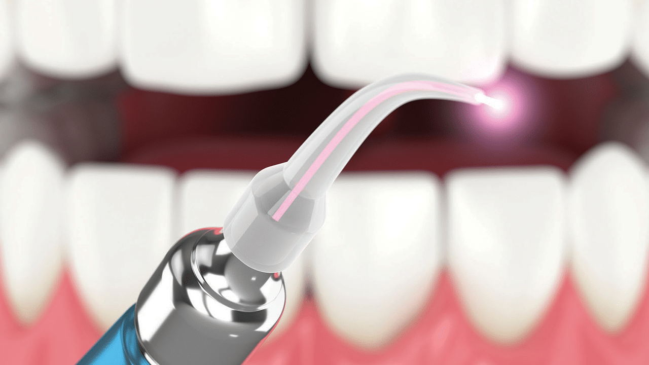
A Closer Look at Laser Safety
Learn how to safely and effectively incorporate diode lasers into dental hygiene practice.
This course was published in the November/December 2026 issue and expires December 2029. The authors have no commercial conflicts of interest to disclose. This 2 credit hour self-study activity is electronically mediated.
AGD Subject Code: 135
EDUCATIONAL OBJECTIVES
After reading this course, the participant should be able to:
- Discuss the dental laser’s mechanism of action.
- Identify best practices to ensure the safe use of the diode laser.
- List contraindications for laser therapy.
The use of laser technology in dental hygiene practice has expanded as clinicians seek minimally invasive options that improve patient comfort and clinical outcomes.1 Diode lasers are commonly incorporated into preventive and therapeutic procedures, including laser bacterial reduction, periodontal therapy, desensitization, management of oral lesions, and additional soft-tissue applications. When used appropriately, lasers may offer benefits such as reduced bleeding, improved healing response, decreased post-operative discomfort, and enhanced clinical precision.1,2 Lasers, however, are regulated medical devices capable of causing serious harm, such as ocular injury, thermal effects, airborne contaminants, and present a fire hazard if safety protocols are not followed.1-3
Dental lasers deliver concentrated light energy that interacts with tissue through absorption, reflection, transmission, or scattering.3 A diode laser is a soft-tissue dental laser that generates energy through a semiconductor and delivers that energy at a specific wavelength. Because diode lasers are primarily absorbed by pigmented tissues and hemoglobin, they are well suited for soft-tissue procedures.1,3
While this technology may enhance clinical effectiveness, it increases the potential for injury if the laser beam is misdirected or safety precautions are ignored. Laser safety protocols must be applied consistently.3
Common Myths About Laser Therapy
Despite the growing incorporation of diode lasers into dental practice, misconceptions persist. Correcting these misunderstandings supports responsible laser use and a safe clinical environment.3,4
Myth 1: Diode lasers are too low-energy to cause harm. Truth: Diode lasers may appear less powerful than surgical or hospital-based lasers, but they are classified as Class IV medical devices, meaning they pose the highest potential risk to health and safety. This designation indicates that diode lasers emit high-energy light capable of causing serious tissue burns, skin injury, and irreversible eye damage if used improperly.1-4 Their ability to penetrate soft tissue and generate thermal effects requires strict adherence to safety protocols. Assuming lasers are inherently safe increases the likelihood of accidental exposure or injury.1-4
Myth 2: Any tinted or dark glasses provide adequate eye protection. Truth: Effective eye protection requires wavelength-specific laser safety eyewear that has been certified according to recognized standards. These glasses are designed to block or absorb the specific wavelength emitted by the laser device, significantly reducing the risk of retinal injury. Generic tinted glasses may reduce visible brightness but do not reliably protect against laser energy. Wavelength-specific eyewear must be available, maintained, and worn consistently by everyone present in the operatory during laser use.
Myth 3: Lasers sterilize everything they touch. Truth: Although diode lasers can reduce the microbial load in treated areas, they do not replace conventional infection control measures. Lasers are an adjunct to care, not a substitute for established protocols.1-4
Myth 4: Low power settings eliminate the need for full safety precautions. Truth: Even when diode lasers are used at lower power settings or for brief procedures, full safety precautions remain mandatory. Reflected or scattered laser energy can still pose a risk to the eyes and skin, and laser plume can still be generated during tissue interaction.4,5 Eye protection, plume evacuation, operatory control, and warning signage must be used consistently when lasers are in use.
Myth 5: Laser safety is only the operator’s responsibility. Truth: Laser safety is a shared responsibility among the entire dental team. While the clinician operating the laser plays a central role, dental assistants, dental hygienists, and other staff members prepare the operatory, monitor patient comfort, enforce protective measures, and limit access to the treatment area.1,5 A team-based approach improves consistency, reduces errors, and reinforces accountability.
Best Practices to Ensure Safety
Eye injury is the most serious laser-related risk and may occur through direct exposure or reflected energy. The retina is particularly vulnerable, and damage may occur without immediate pain or visible symptoms, making prevention critical.3,6
In the dental operatory, the close proximity of the oral cavity to the eyes and the frequent use of reflective instruments increase the risk of accidental exposure. Reflection from mirrors, metal instruments, metal piercings, or other shiny surfaces commonly found in dental operatories can redirect laser energy unpredictably.6 For this reason, eye protection is mandatory whenever the laser is activated, regardless of power settings or treatment duration.
Appropriate laser safety eyewear must be worn by the clinician, patient, dental assistant, and anyone entering the operatory while the laser is in use. Eyewear must be specific to the wavelength and optical density required for the laser being used. The appropriate wavelength designation is printed on the side of the protective glasses. Eyewear should be inspected before each use for scratches, cracks, or pitting, and damaged eyewear must be removed from service. Clinicians should avoid looking directly at the laser beam or reflective surfaces during activation.7,8
Accidental exposure to laser energy can cause burns or unintended tissue damage. These injuries may result from improper aiming, incorrect settings, equipment malfunction, or operator distraction. Controlled hand movements, appropriate settings, and adherence to manufacturer guidelines significantly reduce this risk.8
Clinicians must also be aware of materials used in or around the patient’s lips and oral tissues that may ignite when exposed to laser energy. Petroleum-based products are frequently used by patients and may be flammable when exposed to laser energy.3-5 They should be removed from the treatment area, and nonpetroleum alternatives should be used when moisture or tissue protection is needed. Additionally, alcohol-based disinfectants should not be used while the laser is in operation. Laser tips should only be wiped with dry or water-moistened gauze.
Laser plume is generated when laser energy interacts with tissue, producing smoke that contains carbonized tissue particles, blood fragments, bacteria, viruses, and chemical byproducts.6,7,9 Exposure may irritate the respiratory tract and potentially expose clinicians and patients to infectious or chemical contaminants. Because plume dissipates quickly, risk may be underestimated without adequate evacuation.
High-volume evacuation (HVE) is required during all laser procedures and should be positioned close to the treatment site to capture plume at its source. HVE systems should use tubing with an internal diameter of 8 to 15 mm, which allows for sufficient airflow and suction velocity.4-6,10 Clinicians should wear appropriate masks, such as level 3 or N95 masks. Because plume may contain viable microorganisms and toxic compounds, layered protective strategies are essential.8,11
Oxygen use should be carefully managed. A fire extinguisher must be readily accessible and its location must be known to all clinical personnel prior to initiating laser use.6-8,10 The office should have a designated staff member who develops, implements, and maintains the facility’s fire prevention and evacuation procedures and ensure all staff members are familiar with these protocols.11
When a laser is in use, the operatory is referred to as the nominal hazard zone.8,11 Reflective surfaces should be minimized, and unnecessary instruments removed from the immediate treatment area. Doors should remain closed, and access should be limited during laser procedures. Dental lasers are equipped with safety mechanisms such as key locks, emergency stop buttons, foot pedals, and system timeouts,8,9,11 which should be checked prior to each procedure to confirm proper function.
Contraindications and Precautions
Careful assessment and sound clinical judgment are essential to ensure the safe and effective use of lasers. A thorough review of the patient’s medical history is critical before initiating any laser-assisted treatment. Certain systemic conditions may influence tissue response, healing capacity, or pain perception and should prompt heightened caution.6 Patients with bleeding disorders or those taking anticoagulant medications may be at increased risk for post-operative bleeding, even though diode lasers often promote hemostasis.1-4
Patients who are immunocompromised, including those undergoing chemotherapy, taking immunosuppressive medications, or living with autoimmune conditions, may experience delayed healing or increased susceptibility to infection.11 In these situations, conservative laser settings, meticulous infection control, and close post-operative monitoring are recommended.
Individuals with cardiac conditions, including pacemakers or implantable defibrillators, should also be evaluated carefully.11,12 Although diode lasers generally pose minimal electromagnetic interference risk, consultation with the patient’s physician or device manufacturer may be warranted if uncertainty exists.
Patients with obstructive sleep apnea (OSA) require special consideration during laser-assisted procedures. Supine positioning, mouth opening, and prolonged appointments may compromise airway comfort and tolerance.12,13 Altered oxygenation associated with OSA may affect tissue perfusion and thermal response, potentially increasing sensitivity to heat or delaying tissue recovery. Shorter treatment intervals, careful positioning, conservative energy settings, and continuous assessment of patient comfort are recommended.12
Certain medications may increase tissue responsiveness to light-based therapies. Antibiotics as well as other medications may heighten the risk of exaggerated tissue response or discomfort during laser use.12,13 Clinicians should consider reducing settings, modifying treatment, or deferring therapy.
Patients with autoimmune conditions associated with light sensitivity, such as systemic lupus erythematosus, may have an increased risk of adverse tissue reactions when exposed to laser energy.14 Although diode lasers are not ultraviolet-based devices, heightened photosensitivity and altered inflammatory responses warrant caution. Laser therapy should be evaluated on an individual basis or avoided if risk outweighs benefit.6-8
Patients with neuropathies may have altered pain perception. Reduced sensitivity can mask discomfort or overheating during laser use, increasing the risk of unintended tissue injury.15 Continuous patient communication, conservative energy settings, and careful monitoring are essential. Patients with anxiety disorders or heightened sensitivity to sensory stimuli may require additional explanation, reassurance, or modification of treatment to ensure comfort and cooperation.14
Patients with respiratory conditions may be more sensitive to plume exposure.7,16 In these cases, meticulous plume evacuation is particularly important. If respiratory discomfort occurs during treatment, laser use should be paused or discontinued.
While diode lasers are generally considered safe when used appropriately, elective laser procedures may be deferred during pregnancy as a precautionary measure. When laser treatment is clinically indicated, the lowest effective settings should be selected, and patient comfort, positioning, and stress levels should be carefully monitored.8,11
Laser use should be avoided or modified in areas with suspected malignancy or undiagnosed lesions unless a definitive diagnosis has been established.16 Laser energy may alter tissue appearance and interfere with histologic evaluation.
Active infections accompanied by significant swelling, purulence, or systemic symptoms may require medical management prior to laser intervention.16,17 Areas with thin or fragile tissue, recent surgical sites, or compromised blood supply may be more susceptible to thermal injury.
Special caution is required when working around dental implants. Diode lasers should not be used in direct contact with implant surfaces, as excessive heat may damage the implant–bone interface and compromise osseointegration.15 In dental hygiene practice, laser use around implants is generally limited to conservative bacterial reduction for conditions such as peri-implant mucositis, using low power settings and noninitiated tips.
Laser therapy should be deferred when patient safety cannot be reasonably ensured or when the procedure falls outside the dental hygienist’s scope of practice. In cases of uncertainty, consultation with the supervising dentist or referral to a specialist is the most appropriate course of action. Recognizing contraindications reinforces responsible use.2-4
Accurate and thorough documentation is a critical component of patient safety.4,7 Comprehensive records ensure continuity of care, support clinical decision making, and provide a clear account of how laser therapy was delivered and monitored. Clinical records should include the type of laser used, wavelength, power settings, duration of exposure, use of appropriate protective eyewear for both the patient and clinical staff, and the patient’s response before, during, and after treatment. Any modifications to standard protocols, patient-specific precautions, or interruptions during laser use should also be documented.
By maintaining detailed and consistent documentation, dental hygienists can identify trends, evaluate treatment outcomes, and promptly address adverse reactions or safety concerns.17 This level of documentation supports quality assurance initiatives and helps ensure that laser-assisted care is delivered responsibly and in the best interest of the patient.
Common Safety Errors and How to Prevent Them
Common laser safety errors in clinical practice include improper or inconsistent use of protective eyewear, inadequate plume evacuation, incomplete documentation, failure to post required laser warning signs, and insufficient review or understanding of the patient’s medical history prior to laser use.6-8 Overlooking systemic conditions, medications, or photosensitivity risks may result in inappropriate case selection or adverse tissue responses, compromising patient safety.
Prevention of laser-related adverse events relies on consistent adherence to safety protocols and a proactive approach to risk management.3,4,7 The use of standardized checklists before, during, and after laser procedures promotes consistency and reduces reliance on memory alone. Effective team communication is equally important. Clinicians should feel empowered to pause or discontinue laser use if safety concerns arise, and near-misses should be reported and reviewed to identify opportunities for improvement.4
Laser technology offers valuable benefits in dental hygiene practice, with the potential to enhance patient outcomes, but safe use depends on consistent adherence to established safety principles. Dental hygienists play a central role in protecting patients, colleagues, and themselves through preparation, vigilance, and documentation. When laser safety is fully integrated into daily practice, clinicians can confidently and responsibly utilize this advanced technology.
References
- Abdullah LA, Hashim N, Rehman MM, et al. Effectiveness of diode (810 nm) laser in periodontal parameters and reduction of subgingival bacterial load in periodontitis patients. J Contemp Dent Pract. 2023;24:1008-1015.
- Slot DE, Jorritsma KH, Cobb CM, Van der Weijden GA. The effect of the thermal diode laser (808-980 nm) in non-surgical periodontal therapy: A systematic review and meta-analysis. J Clin Periodontol. 2014;41:681-692.
- Mohammadian M, Jalouti N, Yazdanian M, Keykha E, Hajisadeghi S. Comparison of the effectiveness of diode laser, fluoride varnish, and their combination in treatment of dentin hypersensitivity: A systematic review of randomized clinical trials. Front Oral Health. 2025;6:1550127.
- Porteous MS, Rowe DJ. Adjunctive use of the diode laser in non-surgical periodontal therapy: exploring the controversy. J Dent Hyg. 2014;88:78
- Aimetti M, Mariani GM, Ferrarotti F, Ercoli E, Liu CC, Romano F. Adjunctive efficacy of diode laser in the treatment of peri-implant mucositis with mechanical therapy: A randomized clinical trial. Clin Oral Implants Res. 2019;30:429-438.
- Malcangi G, Patano A, Trilli I, et al. Therapeutic and adverse effects of lasers in dentistry: a systematic review. Photonics. 2023;10(6):650.
- Yahya G, AlAlwi A, Shurayji F, Baroom W, RAjeh M, AbdelAleem N. Comparison of sodium fluoride varnish and diode laser therapy for post-bleaching hypersensitivity: randomized clinical trial. Saudi Dent J. 2022;34:62-67.
- Assaf M, Yilmaz S, Kuru B, Ipci SD, Noyum U, Kadir T. Effect of the diode laser on bacteremia associated with dental ultrasonic scaling: a clinical and microbiological study. Photomed Laser Surg. 2007;25:250-256.
- Moritz A, Schoop U, Goharkhay K, et al. Bacterial reduction in periodontal pockets through irradiation with a diode laser: a controlled clinical study. J Clin Laser Med Surg. 1997;15:33-37.
- Atieh MA, Fadhul I, Shah M, Hannawi H, Alsabeeha NHM. Diode laser as an adjunctive treatment for peri-implant mucositis: systematic review and meta-analysis. Int Dent J. 2022;72(6):735-745.
- Saglam M, Kantarci A, Dundar N, et al. Clinical and biochemical effects of diode laser as an adjunct to nonsurgical treatment of chronic periodontitis: a randomized controlled clinical trial. Lasers Med Sci. 2014;29:37-46.
- Dukić W, Bago I, Aurer A, Roguljić M. Clinical effectiveness of diode laser therapy as an adjunct to non-surgical periodontal treatment: a randomized clinical study. J Periodontol. 2013;84:1111-1117.
- Üstün K, Erciyas K, Sezer U, et al. Clinical and biochemical effects of 810 nm diode laser as an adjunct to periodontal therapy: a randomized split-mouth clinical trial. Photomed Laser Surg. 2014;32:61-66.
- Borrajo JL, Varela LG, Castro GL, Rodríguez-Nuñez I, Torreira MG. Diode laser (980 nm) as adjunct to scaling and root planing. Photomed Laser Surg. 2004;22:509-512.
- Qadri T, Tunér J, Gustafsson A. The short-term effects of low-level lasers as adjunct therapy in the treatment of periodontal inflammation. J Clin Periodontol. 2005;32:714-719.
- Carpino G, et al. Diode laser adjunctive to non-surgical periodontal therapy reduces matrix metalloproteinase-8 levels and enhances clinical outcomes: a clinical and biochemical study. Open Dent J. 2018;12:281-291
- Üstün K, Erciyas K, Sezer U. Clinical and biochemical effects of 810 nm diode laser as an adjunct to periodontal therapy: a randomized split-mouth clinical trial. Photomed Laser Surg. 2014;32:61-66.
From Dimensions of Dental Hygiene. November/December 2026; 24(6):32-35