

The Rare Disorder Hiding in Plain Sight
Pachyonychia congenita can cause distinctive changes to the nails, skin, and oral tissues, making oral health professionals essential to recognizing this debilitating disorder.
This course was published in the November/December 2026 issue and expires December 2029. The authors have no commercial conflicts of interest to disclose. This 2 credit hour self-study activity is electronically mediated.
AGD Subject Code: 730
EDUCATIONAL OBJECTIVES
After reading this course, the participant should be able to:
- Define pachyonychia congenita (PC).
- Identify the physical and oral manifestations of PC.
- Discuss the appropriate management of patients with PC.
Caused by mutations of the genes responsible for producing keratin, pachyonychia congenita (PC) is a rare and debilitating skin disorder with unusual oral manifestations.1 Keratin consists of proteins naturally produced by the body to help support strong hair, skin, and nails. Individuals with PC, however, experience an alteration of keratin genes that give rise to ectodermal structures, such as the skin and oral mucosa.
According to the Pachyonychia Congenita Project International PC Research Registry, as of June 2026, there are 1,322 genetically confirmed cases worldwide, with nearly half presenting in the United States. California, New York, Florida, and Texas have the most cases, respectively, and confirmed cases have been diagnosed in nearly all 50 states and the District of Columbia.1
Considered an autosomal dominant disorder in which only one copy of the mutated gene is needed to manifest clinically, PC is found in both men and women. Interestingly, 30% to 40% of PC cases occur in individuals with no previous family history.2 Generally, PC does not decrease life expectancy; however, many patients with PC experience chronic pain that negatively impacts quality of life. Due to the likely involvement of the oral tissues, oral health professionals should be knowledgeable about PC and its impact on patient care.
Signs and symptoms of PC typically begin in early infancy or early childhood and vary according to the genes involved (Table 1). Thickened discolored fingernails and toenails; painful hyperkeratosis on the palms of the hands and soles of the feet; oral leukokeratosis involving the tongue, throat, and buccal and labial mucosa; persistent hoarseness; and feeding difficulties are commonly seen in patients with PC.
| Physical |
|---|
|
| Oral/Dental |
|
Additionally, epidermal cysts, severe temporary ear pain, and excess ear wax have all been noted in patients with PC. Angular cheilitis, natal teeth, and dental anomalies, such as enamel loss, premature decay, abnormal tooth morphology, supernumerary teeth, and anodontia, have also been observed in certain phenotypes.2-6
Physical Manifestations
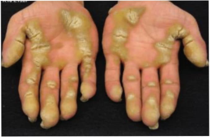
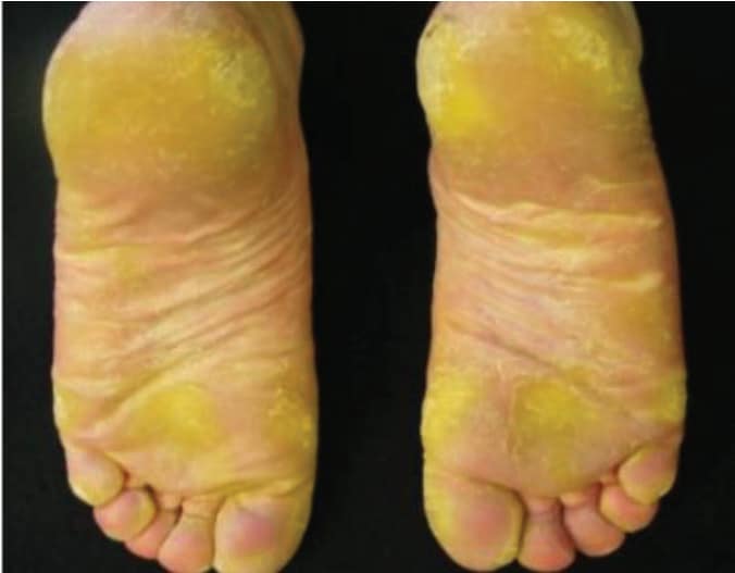
The main clinical features of PC are hypertrophic nail dystrophy involving fingers and toes and painful palmoplantar hyperkeratosis (excessive thickening of skin on hands and feet), often accompanied by blistering (Figures 1 and 2). These three symptoms are considered the triad of PC.4 Additionally, PC causes various types of lesions and cysts involving skin and hair, and palmoplantar hyperhidrosis (excessive sweating of hands and feet).3-6
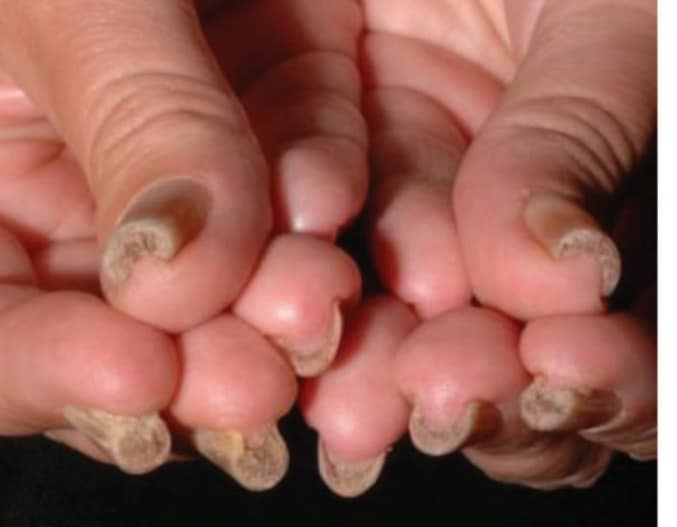
Most patients affected by PC develop nail dystrophy (Figure 3). This condition is characterized by thickened and distorted nails, that typically appear during the first years of life. Affected nails tend to be discolored and elevated from the nail bed due to subungual hyperkeratosis. Samuelov et al3 found that out of 815 individuals with PC, 75% had fingernail involvement and 96% had toenail involvement.
Interestingly, PC does not always result in dystrophy of all nails. Individual or multiple nails may be affected.4,6-8 A case report of an infant girl discussed unilateral involvement of fingernails. The nails of the left hand and foot were barely affected with only minor involvement; however, both the right hand and foot exhibited hypertrophic dystrophy of all nails.9
Along with nail dystrophy, hyperkeratosis of the palms of the hands is present in some genetic manifestations. Hyperkeratosis of the soles of the feet is present in all gene variations of PC. Furthermore, pain associated with plantar hyperkeratosis is also present in each subtype, with as high as 100% of patients reporting pain.4 Plantar pain is considered the most debilitating symptom of PC, often causing patients to use crutches, canes, and wheelchairs and seek medical and/or surgical interventions.4,5
Oral Manifestations
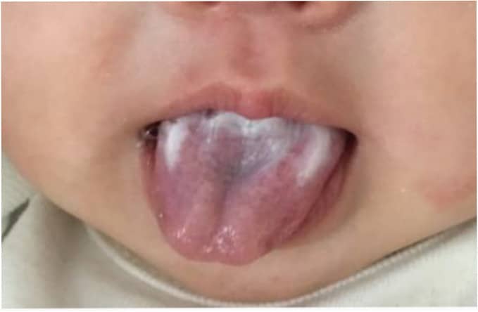
In addition to the physical characteristics of PC, oral manifestations are common. Oral leukokeratosis of the buccal mucosa, tongue, and throat is very common in patients with PC of all ages (Figure 4). Feeding difficulties in infancy, the presence of natal teeth (teeth present at birth),3,4,10,11 and partial and complete anodontia have been documented.6 Median rhomboid glossitis, angular cheilitis, hoarseness and laryngeal abnormalities, breathing difficulties, stridor (loud or noisy breathing), and laryngeal obstruction have all been noted in cases of PC.3,4,6,12-14 Additionally, first bite syndrome (FBS), a condition that causes ear pain associated with salivation and taking the first bite of food, has been seen in cases of PC.5
One of the most common manifestations of PC, oral leukokeratosis has been reported in patients who present with mutations in any of the five PC-associated keratin genes. Samuelov et al3 noted that oral leukokeratosis was reported in 440 of 812 patients. Of those individuals affected, 60% reported the first signs of oral leukokeratosis prior to age 1 with the most common area affected being the buccal mucosa. This study goes on to discuss the positive correlation between oral leukokeratosis, nursing difficulties, hoarseness, and the use of walking aids due to pain associated with plantar hyperkeratosis.
A case series published by Goldberg et al5 detailed the mucosal involvement in nine children with PC. Each had oral leukokeratosis and several had hoarseness with vocal cord involvement and difficulties with feeding accompanied by failure to thrive.
McCarthy et al4 reported in rare cases when oral leukokeratosis extends to the larynx, breathing obstruction can create a life-threatening situation. Close monitoring of patients, especially infants, who experience hoarseness must occur. Persistent hoarseness could be a sign of respiratory distress and careful examination by an otolaryngologist is necessary. Interestingly, oral leukokeratosis may present similarly to Candida albicans and can be misdiagnosed and treated as such.4 Therefore, a thorough health history is needed to rule out similar conditions.
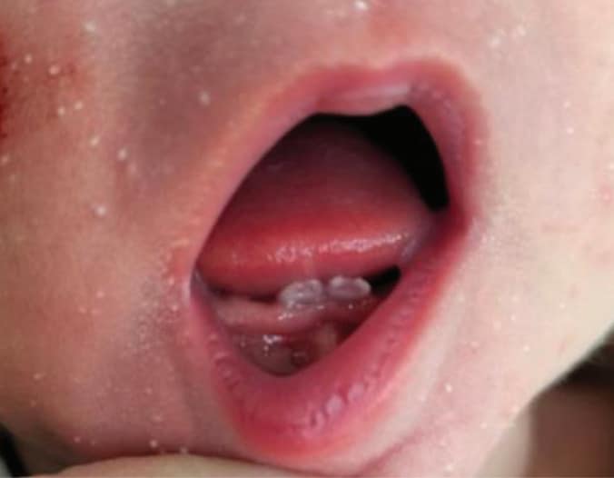
The presence of natal teeth is another unusual finding seen in patients with PC (Figure 5). Dabbagh et al11 reported on a newborn boy with PC who had 12 malformed natal teeth, six on the maxilla and six on the mandible, and fluid-filled lesions on the alveolar crest.12 The natal teeth were loose and subsequently removed to prevent aspiration and nursing difficulties.
A case study by Youness et al15 discussed a 3-month-old infant with PC who had dystrophic nails, hyperhidrosis, and two natal teeth on the lower arch. While oral leukokeratosis is common, newborns with natal teeth are somewhat less so. However, natal teeth may hinder breastfeeding and may inhibit an infant’s growth.3
Median rhomboid glossitis (MRG) has also been observed in cases of PC. While not widely documented, MRG was found in a 3-year-old girl who had a lesion on the dorsum of the tongue anterior to the circumvallate papillae that appeared denuded and rhomboidal in shape.13 Nystatin was prescribed and the lesion subsided somewhat, but did not completely resolve. The authors of the case study went on to speculate that, in this instance, because the nystatin did not resolve lesion totally, the MRG was most likely caused by a C. albicans infection and PC, not just C. albicans alone.13
FBS is a rare condition that occurs in patients with PC. FBS causes intense pain upon taking the first bite or swallow of something. The pain occurs in the area around the ears and temporomandibular joint, can be unilateral or bilateral, and is thought to be associated with an impairment of the sympathetic innervation in the parotid gland.14 The pain is intense but typically diminishes after a short time.16
FBS is more common in babies and younger children and may cause feeding difficulties and failure to thrive. This condition is usually resolved as individuals age.1 Goldberg et al5 describe an infant with PC and FBS who exhibited strong crying, fussiness, and ear-pulling at the beginning of every feeding. Ultimately, a feeding tube was placed from 8 to 11 months of age because of her failure to thrive. FBS is not only a symptom of PC but can be seen in patients with tumors in the head or neck region, who have undergone surgical procedures in the head or neck region, or had cancer treatment for head/neck tumors, among other reasons.5
Genetic Counseling
PC may be diagnosed based on clinical appearance, however, a definitive diagnosis can only be obtained through genetic testing. Genetic testing provides a complete diagnosis, including the type of PC and specific mutation a patient may have. Molecular genetic testing approaches can include serial single-gene testing, multigene panel, and more comprehensive genomic testing.17
Multigene panel testing includes KRT6A, KRT6B, KRT6C, KRT16, KRT17, and other genes of interest. The genes included in the panel and the diagnostic sensitivity of the testing used for each gene varies by laboratory and are likely to change over time. In some laboratories, panel options may include a custom laboratory-designed panel and/or tailored phenotype-focused exome analysis that includes genes specified by the clinician. The methods used in a panel may include sequence analysis, deletion/duplication analysis, and/or other nonsequencing-based tests.17 More comprehensive genomic testing includes exome sequencing and genome sequencing. These tests provide detailed information on the mutation of a different gene or a gene that expresses a similar clinical presentation.17
Genetic counseling that includes family history and genetic testing plays an important role in family planning and risk assessment.17 Genetic counseling provides individuals and families with information on the mode of a disease’s inheritance and implication of genetic disorders. This knowledge can then empower patients and families to make informed medical and personal decisions. PC is inherited as an autosomal dominant pattern with a 50% chance of inheriting the disorder. Interestingly, approximately 30% of PC cases appear to be caused by a de novo pathogenic variant, which is a gene variant that occurs spontaneously and is not inherited.18
Clinical Diagnosis and Treatment
Clinical diagnostic criteria for PC include the triad of toenail thickening, plantar keratoderma, and plantar pain, which is present in 97% of individuals with genetically confirmed PC by age 10.19 No specific treatment for PC is currently available. Patients with PC need a personalized and combined approach using medical and surgical procedures as well as pharmacotherapy. Additional research on improved and targeted treatment options for patients with PC is necessary.
Oral Considerations and Management
PC’s most common oral manifestation is oral leukokeratosis. In some cases, it is present very soon after birth and may be the first sign of PC. Oral leukokeratosis often appears as thickened yellowish-white patches on the tongue, lips, and buccal mucosa. This condition is often misdiagnosed as C. albicans and an antifungal is prescribed. However, if treatment for candidiasis is not effective, thorough examination of the nails should be performed and PC should be considered as a possible diagnosis.3,5
In some cases, leukokeratosis is found on laryngeal surfaces, which may cause hoarseness and/or stridor. In infants, extreme cases of oral leukokeratosis may cause feeding and breathing difficulties, and even death.4,5 Oral leukokeratosis may also be confused with other white lesions (leukoplakias) of the oral cavity, such as lichen planus or leukoedema, and a biopsy may be needed to determine the exact histological analysis of the tissue.1
Careful monitoring by the patient’s pediatrician and other specialists, such as an otolaryngologist, is critical. As the child ages, gentle brushing of the tongue with a soft or toothbrush or using a tongue scraper can be suggested. Additionally, a mouthrinse can be recommended if the oral tissues are irritated. Many over-the-counter and prescription options are available for oral discomfort.
Angular cheilitis, inflammation and fissuring at the commissures of the mouth, is also a frequent finding in patients with PC. Treatment may include a topical antifungal, lip balm, or petroleum jelly to keep the corners of the mouth protected; a nutritious diet with foods high in iron, vitamin B, and protein; and proper hydration. Furthermore, an oral or topical antibiotic can be prescribed if the infection is deemed bacterial rather than fungal.20
Other oral manifestations of PC, such as natal teeth, enamel hypoplasia, abnormal tooth morphology, and supernumerary teeth, should be dealt with in consultation with the patient’s physician and possibly other medical and oral health professionals to manage the complicated symptoms and manifestations of this unusual disorder. Thus, a thorough medical and dental history should be obtained to determine the best way to manage a patient with this disorder and the variety of symptoms it presents.
Conclusion
PC is a rare disorder of the genes that produce keratin. PC is classified based on the keratin gene involved. PC presents with a variety of nail and skin conditions, as well as an array of oral manifestations including oral leukokeratosis, angular cheilitis, natal teeth, median rhomboid glossitis, and FBS. As cases grow across the US and the world, oral health professionals should be aware of the debilitating oral and physical symptoms of this unusual disorder and be prepared to properly manage patients with this challenging condition. A multidisciplinary approach to the myriad symptoms of PC is integral in effective patient management.
References
- Pachyonychia Congenita Project. Connecting and Helping Patients. Available at pachyonychia.org/pc-data. Accessed September 20, 2026.
- MedlinePlus. Pachyonychia congenita. Available at medlineplus.gov/genetics/condition/pachyonychia-congenita/#inheritance. Accessed September 20, 2026.
- Samuelov L, Smith FJD, Hansen CD, Sprecher E. Revisiting pachyonychia congenita: a case‐cohort study of 815 patients. Br J Dermatol. 2020;182:738-746.
- McCarthy R, de Brito M, O’Toole E. Pachyonychia congenita: clinical features and future treatments. Keio J Med. 2025;74:52-60.
- Goldberg I, Mashiah J, Kutz A, et al. Symptomatic mucosal involvement in pachyonychia congenita: challenges in infants and young children. Br J Dermatol. 2020;182:708-713.
- Gönül M, Gül Ü, Kılıç A, Soylu S, Koçak O, Demiriz M. A case of pachyonychia congenita with unusual manifestations: an unusual type or a new syndrome? Int J Dermatol. 2015;54:334-337.
- Bhengra MP, Choudhary R, Kumar P, Chaudhary SS. A familial case of pachyonychia congenita. Journal of Pakistan Association of Dermatologists. 2015;25(1):62-65.
- Rathore PK, Khullar V, Das A. Pachyonychia congenita type 1: case report and review of the literature. Ind J Dermatol. 2016;61:196-199.
- Forrest C, Casey G, Mordaunt DA, Thompson EM, Gordon L. Pachyonychia congenita: a spectrum of krt6a mutations in Australian patients. Pediatr Dermatol. 2016;33:337-342.
- Azkur D, Erkoçoğlu M, Civelek E, Ünite GE, Kocabaş CN. A rare genetic disease: pachyonychia congenita type 2. Turkish Journal of Pediatric Diseases. 2013; 4: 193-195.
- Dabbagh B, Cukier O, Yeganeh M, Halal F, Ferraz dos Santos B. Pachyonychia Congenita Associated with a Novel Variant of KRT17 Presenting Unusual Oral Manifestations. Journal of Dentistry for Children. 2019;86(1):61-63. Accessed May 27, 2026. https://research-ebsco-com.eps.cc.ysu.edu/plink/21a16f6f-2df3-3a3d-a7aa-a37c075e0cc2
- Ceyhan AM, Yildirim M, Akkaya VB, Yasan H. Persistent hoarseness in a patient with pachyonychia congenita: an early sign of laryngeal involvement. Int J Dermatol. 2009;48:1346-1348.
- Karen J, Schaffer J. Pachyonychia congenita associated with median rhomboid glossitis. Dermatol Online J. 2007;13:21.
- Barati B, Ghazizadeh M, Shahzamani A. Successful management of an idiopathic first bite syndrome: A case report and review. Clin Case Rep. 2024;12:e8880.
- Youness Z, Hallal M, Makhoul R, Bassukas ID. A newly documented rare case of pachyonychia congenita ii in a three‐month‐old baby. Case Reports in Dermatological Medicine. 2025;2025:1-4.
- National Institute of Arthritis and Musculoskeletal and Skin Diseases. Pachyonychia Congenita. Available at niams.nih.gov/health-topics/pachyonychia-congenita. Accessed September 20, 2026.
- Smith FJD, Hansen CD, Hull PR, et al. Pachyonychia congenita. GeneReviews®. Available at ncbi.nlm.nih.gov/books/NBK1280. Accesssed September 20, 2026.
- Wilson NJ, Leachman SA, Hansen CD, et al. A large mutational study in pachyonychia congenita. J Invest Dermatol. 2011;131:1018-1024.
- Eliason MJ, Leachman SA, Feng B, Schwartz ME, Hansen CD. A review of the clinical phenotype of 254 patients with genetically confirmed pachyonychia congenita. J Am Acad Dermatol. 2012;67:680-686.
- Cleveland Clinic. Angular Cheilitis. Available at https://my.clevelandclinic.org/health/diseases/21470-angular-cheilitis. Accessed September 20, 2026.
From Dimensions of Dental Hygiene. November/December 2026; 24(6):28-31