Why the Dual-Scan Protocol Sets Your Implant Plan Up for Success
The dual-scan protocol takes the guesswork out of implant planning by pairing CBCT data with a well-adapted diagnostic denture. When executed correctly, it streamlines data integration and ensures implant placement follows a fully prosthetically driven approach.
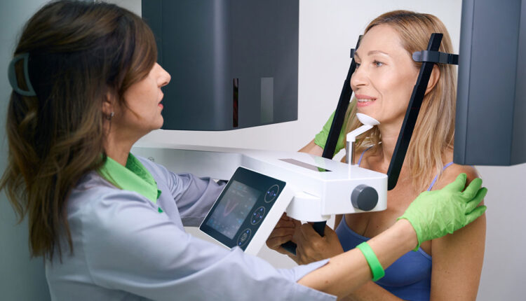
Before implant planning can reach its full digital potential, clinicians must capture precise cone-beam computed tomography (CBCT) data anchored by a stable, well-adapted diagnostic denture. Once the ideal tooth position, vertical occlusal dimension (VOD) in centric relation (CR) and prosthesis design have been established, the next step is to obtain a CBCT scan. If a diagnostic denture is available, the dual-scan protocol becomes an essential technique.1
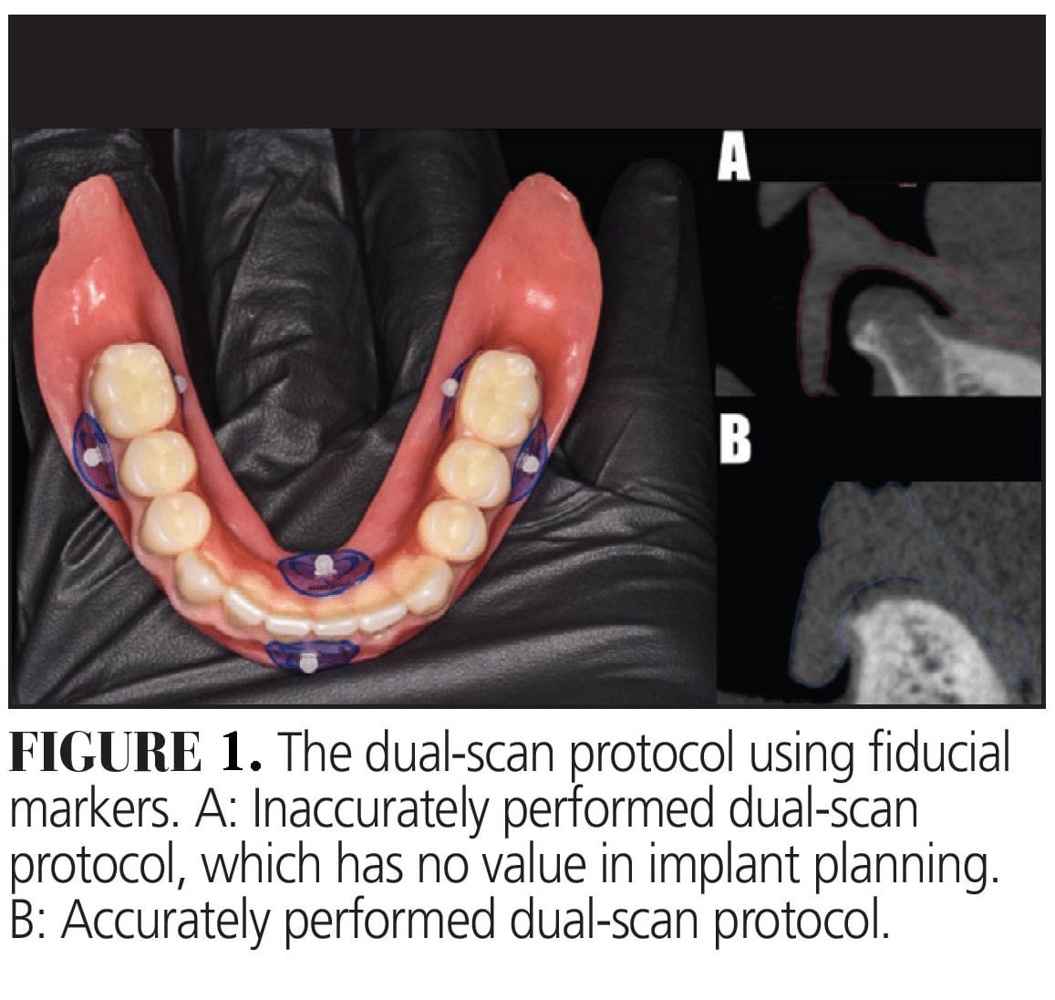
This protocol involves acquiring two CBCT scans: one with the patient wearing the diagnostic denture and another of the denture alone, while incorporating five to six fiducial markers on the denture (with a minimum of three). Two critical considerations in the dual-scan procedure are proper soft tissue adaptation and stable occlusion of the diagnostic denture.2 Inadequate adaptation of the denture base to the soft tissue or an unstable occlusal relationship can result in errors that mislead the planning process (Figure 1). Therefore, ensuring accuracy during the first diagnostic phase is paramount, as it serves as the foundation for the entire workflow.
 A well-adapted and functionally stable diagnostic denture enhances the reliability of data integration in implant simulation software, allowing for a truly prosthetically driven treatment plan that optimizes implant positioning while maintaining biological, functional, and esthetic considerations.2 When using a digital wax-up for terminal dentition cases, the CBCT scan should be acquired with the two jaws separated.3 This separation ensures a clear distinction between the maxillary and mandibular arches, facilitating the registration process between STL and DICOM data. Unlike the dual-scan protocol used with diagnostic dentures in which the scan is taken in closed occlusion to capture the established tooth position and restorative space, the open-jaw CBCT approach prevents overlapping structures and enhances the accuracy of digital file alignment.3
By prioritizing accuracy in the early scanning phase, clinicians set the stage for predictable, prosthetically guided implant placement and smoother workflows from start to finish.
References
- Resnik R. Dual CBCT scanning technique for completely edentulous arches. Dentistry Today. 2016;35(12):50-53.
- Moura GF, Siqueira R, Meirelles L, Maska B, Wang HL, Mendonça G. Denture scanning technique for computer-guided implant-supported restoration treatment of edentulous patients. J Prosthet Dent. 2021;125:726-731.
- Papaspyridakos P, Bedrossian A, De Souza A,Bokhary A, Gonzaga L, Chochlidakis K. Digital workflow in implant treatment planning for terminal dentition patients. J Prosthodont. 2022;31:543-548.
This originally appeared in Nagai T, AlQallaf H. Precision planning in full-arch implant rehabilitation. Decisions in Dentistry. 2025;11(4):32-35.
