Mastering Maintenance for Long-Term Implant Success
Effective peri-implant maintenance therapy is essential for preventing biological and mechanical complications and ensuring the long-term success of dental implants.
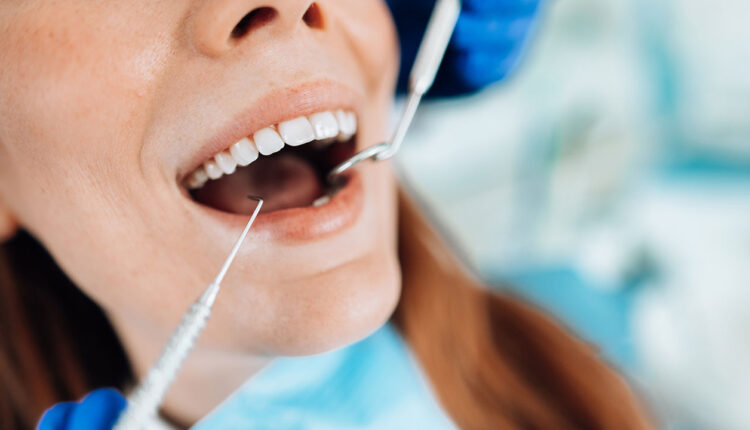
Peri-implant maintenance therapy includes assessment of prosthetic stability, occlusion, presence of interproximal contact, examination of peri-implant hard and soft tissues, professional debridement, and review of oral hygiene instructions.
Prosthetic stability is directly related to the tightness and integrity of the abutment and abutment screw. Verification of prosthesis stability can be done by applying lateral and vertical pressure to the restoration and confirming no movement is present. Confirmation of a protective occlusal scheme can be done using a piece of articulating paper and shimstock.
Evaluation of interproximal contact can be done using dental floss. Interproximal contact loss (ICL) between an implant restoration and an adjacent tooth is quite common, with a prevalence of ~50% and occurring most frequently on the mesial surface.1-3 ICL may be caused by the natural mesial tooth migration compensating for wear, progressive growth of the dento-alveolar complex, and the location and size of the contact itself. ICL leads to food impaction, which is associated with worsened periodontal parameters (higher plaque index, gingival index, and probing depths) and caries formation.2,3 As a result, peri-implant disease is more common at sites with an open interproximal contact.
Clinical examination of the peri-implant soft tissues includes assessment of the amount and quality of tissue, presence of inflammation, and gentle probing. Several studies show that inadequate keratinized tissue and thin peri-implant tissue result in higher bleeding on probing, plaque accumulation, gingival recession, and possibly crestal bone loss over time.4,5 Probing around implants is recommended at each maintenance appointment to determine if probing depths have increased. Early detection and treatment of peri-implant mucositis are important to prevent peri-implantitis.
Radiographic examination of peri-implant hard tissues includes a periapical and vertical bitewing taken annually. This aids with assessing whether progressive bone loss has occurred.
Nonsurgical debridement may include the use of ultrasonic devices, curets, air-abrasive devices, lasers, and local antimicrobial agents. Reviewing oral hygiene instruction with the patient is critical and should be done at each maintenance appointment.
Screw-retained restorations have the advantage of retrievability; however, mechanical complications are common and they are not immune to the biological complication of peri-implant disease. Early detection and correction of these complications directly affect the long-term success and survival of the restoration and implant.
References
- Varthis S, Randi A, Tarnow DP. Prevalence of interproximal open contacts between single-implant restorations and adjacent teeth. Int J Oral Maxillofac Implants. 2016;31:1089-1092.
- Gasser TJW, Papageorgiou SN, et al. Interproximal contact loss at implant sites: a retrospective clinical study with a 10-year follow-up. Clin Oral Implants Res. 2022;33:482-491.
- Varthis S, Tarnow DP, Randi A. Interproximal open contacts between implant restorations and adjacent teeth. Prevalence-Causes-Possible solutions. J Prosthodont. 2019;28:806-810.
- Suarez-Lopez Del Amo F, Lin G-H, et al. Influence of soft tissue thickness on peri-implant marginal bone loss: a systematic review and meta-analysis. J Periodontol. 2016; 87:690-699.
- Lin GH, Chan HL, Wang HL. The significance of keratinized mucosa on implant health: a systematic review. J Periodontol. 2013;84:1755-1767.
This information originally appeared in Casarez-Quintana AR. Enhancing long-term success with screw-retained implants. Decisions in Dentistry. 2024; 10(5):10-14
