A Hidden Stroke Risk Revealed on a Panoramic X-Ray
A case report links a calcified stylohyoid ligament to carotid artery plaque, highlighting a potential stroke risk visible on dental radiographs. Oral health professionals are uniquely positioned to identify this silent threat before serious complications occur.
A recent case report in the International Clinical and Medical Case Reports Journal sheds light on a critical, yet often overlooked role dentists play in preventing strokes. The report details a case of vascular Eagle syndrome, in which a calcified stylohyoid ligament was found to be pressing against the internal carotid artery, potentially contributing to the formation of plaque and increasing stroke risk.
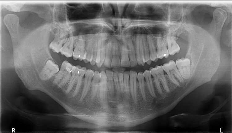
The calcified ligament was visible on a routine panoramic radiograph. Unlike other medical settings, oral health professionals are often the only healthcare providers capturing this specific view, putting them on the front lines of identifying this anatomical anomaly.
Although most cases of elongated styloid processes or calcified stylohyoid ligaments are asymptomatic, their proximity to major vascular structures can, in rare cases, lead to serious complications, such as carotid dissection or stroke. In this report, the patient had already begun developing plaque on the artery adjacent to the ossified ligament; plaque that was not present on the opposite side.
This case emphasizes the importance of carefully examining panoramic images not just for dental concerns but for broader anatomical clues that could signal systemic risk. Click here to read the case study.
