Investigating Probe Marking Degradation and Its Impact on Implant Health
A study was conducted to examine how cleaning, sterilization, and frictional wear affect the color markings on dental probes and their impact on implant surfaces. Results showed that while sterilization didn’t degrade the color markers, friction from titanium-based implants and abutments led to visible wear on the probes and color transfer, with varying degrees of impact depending on the material.
In order to ascertain how probe markings degrade and the effects of color degradation on implant health, a study was initiated. The purpose of this study was to answer the following questions:
- Do the color markers degrade as a result of cleaning and sterilization?
- Is the colored paint removed during the action of probing the implant or abutment?
- What is the force required for transfer of colored material onto different implant and abutment materials compared to clinically recommended peri-implant probing force?
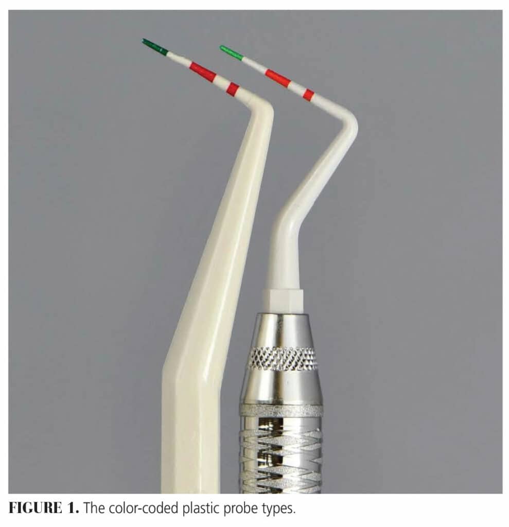
Twenty new, unused plastic probes from two manufacturers (A and B) were selected for the study. Both sets of probes had similar proprietary color code graduations of green and red (Figure 1).
 To assess if the color markings were being removed during the cleaning and sterilization processes, one probe from each manufacturer was chosen for a preliminary study. A standardized photographic setup was developed with the probe placed a specific distance from the camera.
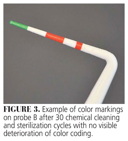
Photos of the probes prior to cleaning and sterilization were taken. The cleaning sequence involved wiping the probes with isopropanol disinfection cloths and then placing them in sterilization pouches. The probes were heat sterilized following the manufacturer’s protocol. After every five cycles, images of the probes were taken, and visual examination was conducted. This process was repeated for a total of 30 cycles.
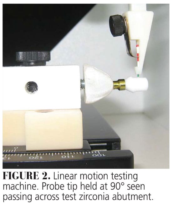
The remaining 18 colored probes, nine from each manufacturer A and nine from manufacturer B, were used to investigate if the color markings faded due to frictional wear between the implant or implant abutment during use. To control the force applied at the probe tip, a custom linear movement test probing model was developed. The sample implants and abutments were placed at a 90° angle to a clamp, which held the test plastic probe tip (Figure 2). The probe tips were then moved across the surfaces of four different implant types and five abutments in single, one-direction passes. The initial weight on the probe clamp was fixed at 10 g equivalent to a force of approximately 0.1 N.
 Of the four implant types, one was made from zirconia, two were titanium grade 4, and one was titanium grade 5 alloy. The five abutment materials used were: zirconia, titanium nitride, titanium aluminum vanadium, titanium aluminum niobium, and gold alloy. Images (1:1 magnification) of the probes, implants, and abutments were obtained prior to testing to serve as controls.
Each probe was moved across the surface of either the implant or the abutment, and after each pass, photographs were taken. Testing stopped when the green color was visibly transferred to the implant or abutment, or when the green color was removed from the probe tip, exposing the underlying white plastic core. For further analysis, select implants and abutments were viewed using a scanning electron microscope (SEM).
Results
In the cleaning and sterilization study, no observable color alterations occurred on the probe tips even after 30 cycles. Both the A and B probes appeared identical to the new untouched control (Figure 3).
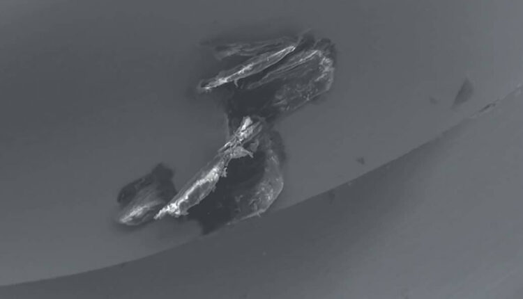
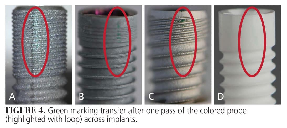
 Color loss from the tips did occur when the probes passed across the implant materials. The titanium-based implants showed visible deposits of green debris. This occurred after just one pass with a force of 0.1 N on the probe tip (Figure 4). Although the green debris was present on the zirconia implant, it was relatively more challenging to detect due to the smoother, reflective surface (Figure 4D).
We also assessed the number of passes — made by the probe at 0.1 N across the implants — needed to remove the green paint and expose the underlying plastic core. The plastic base was evident with the three titanium implants after two passes from either probe A or probe B, while the zirconia implant required five passes for probe B and six for probe A (Figure 4). This was an indicator of how much colored paint was being removed during each probing.
 The abutments exhibited varying results. The locator abutment with a titanium nitride, gold-colored coating exhibited green color on a single pass with both probe types (Figure 5A). The underlying probe core was exposed at 12 passes for probe A and eight for probe B. For the gold alloy abutment, probe A resulted in green debris after the first pass, with the probe’s core exposed after eight passes. Probe B showed green debris deposits on the second pass and exposed the core after 18 passes (Figure 5B).
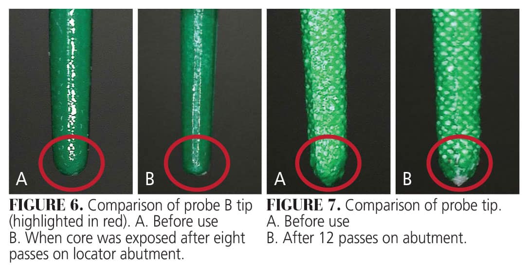
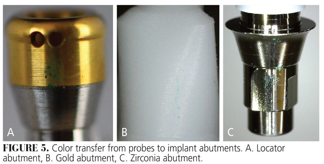 The zirconia abutment displayed green debris with a single pass of either probe type, at 0.1 N, consistent with the zirconia implant (Figure 5C). Additionally, the tip of probe A exposed the probe’s core material after eight passes over the abutment surface, whereas probe B required 10 passes (Figure 6A-B and Figure 7A-B).
 In contrast, the titanium aluminum vanadium and titanium aluminum niobium alloy abutments did not display any visible debris on their surfaces, even after 30 passes. This was mainly due to the difficulty in visualizing green paint on the smooth metal surface. The green paint did not adhere to the titanium alloy’s smooth surface in contrast to the other materials. However, both probe tips showed visible wear exposing the probe’s core after 30 tests.
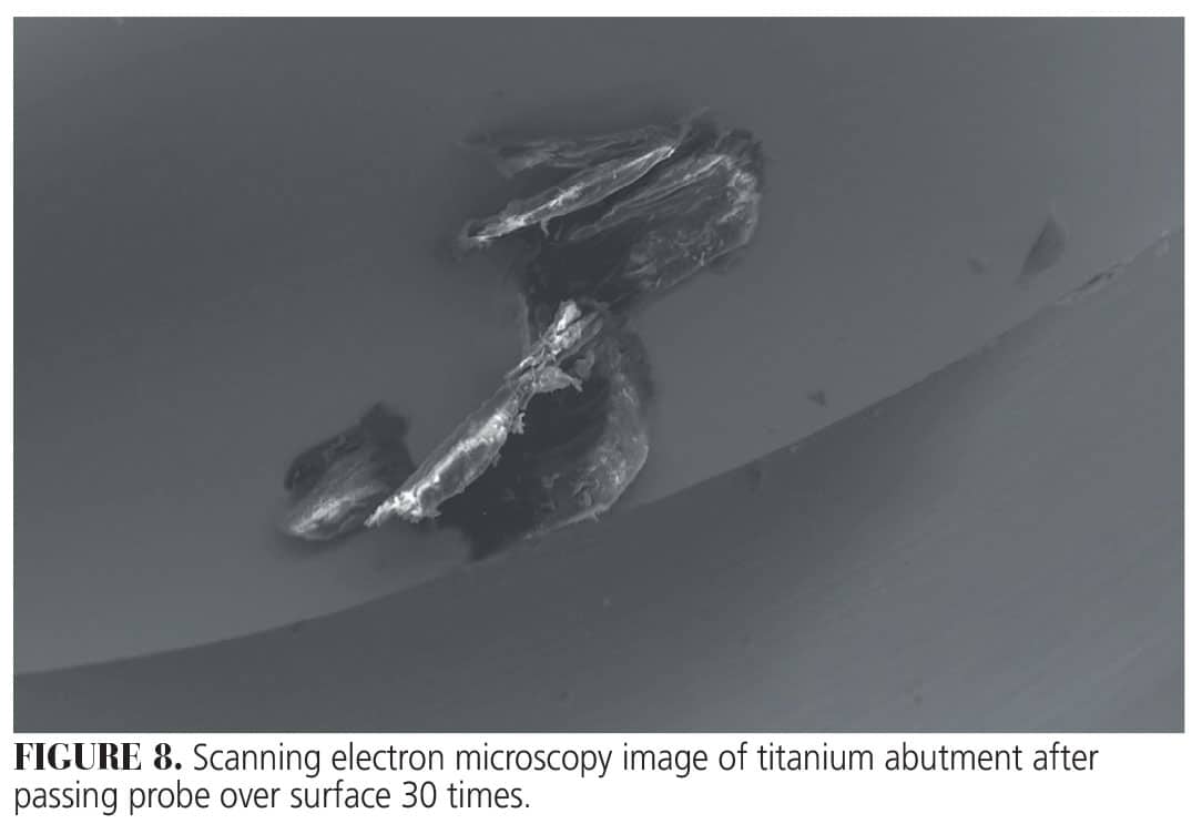 To better understand the surface alterations on the titanium abutments in particular, SEM images were captured. They showed vertical streaks and debris present after 30 passes (Figure 8).
This information originally appeared in Kim SJ, Wadhwani C, Ferracane J, O’Brien R, Katancik JA. The impact of dental probe wear on implant health. Decisions in Dentistry. 2024;10(4):32-35.
