Enhancing Diagnostics and Precision Care With CBCT
Cone-beam computed tomography is transforming periodontal diagnostics by providing highly detailed imaging of bony defects, root morphology, and periodontal-endodontic pathology. This advanced technology not only aids in precise treatment planning but also elevates clinical success rates and patient outcomes.
In the evolving field of periodontics, cone-beam computed tomography (CBCT) has emerged as a powerful tool for enhancing diagnostic accuracy and treatment precision. From evaluating complex periodontal defects to detecting root anomalies and combined periodontal-endodontic lesions, CBCT offers unparalleled imaging detail. By integrating this technology into clinical practice, dental professionals can improve patient care, optimize treatment outcomes, and navigate challenging cases with greater confidence. This article delves into the critical applications of CBCT in periodontics, shedding light on its impact on regenerative therapy and interdisciplinary collaboration.
Common locations of localized advanced periodontitis and its bone destruction frequently include interproximal and furcation areas, and these defects are often candidates for regenerative periodontal procedures.1 Cone beam scans provide the ability to evaluate the configuration of these defects, aiding in the diagnosis of their severity and extent. It has been shown that CBCT yields the highest sensitivity and greatest diagnostic accuracy for the detection of these periodontal defects.2 The number of remaining bony walls of the defect often dictates the extent of the periodontal regeneration attainable. With this information, the later success of periodontal regenerative therapy may be made clearer from the CBCT data.
Similarly, CBCT scans can help identify complicating root morphology, such as the presence of developmental root grooves. These, in turn, predispose the tooth to periodontal attachment loss. Developmental grooves most frequently present on maxillary incisors; more specifically, the central incisors (0.28%) and lateral incisors (4.4%).3,4 These grooves are often associated with soft tissue attachment loss and the progression of bacteria subgingivally.4 With prior knowledge of the configuration and extent of the developmental groove, the prognosis of the tooth — along with the success rate of periodontal therapy for tooth retention — can be determined.
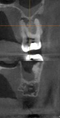
Lastly, from an interdisciplinary perspective, the ability to visualize circumferentially around a tooth with cone beam imaging can help clinicians identify the presence of combined periodontal-endodontic pathology.5 In fact, the literature indicates that peri-radicular pathology can be detected with greater accuracy from CBCT scans than it can from conventional, two-dimensional radiography (Figure 1).6 The impact from this increased accuracy in the diagnosis of periodontal-endodontic lesions translates to more precise treatment planning, improved clinical success, and better overall patient care.
Cone-beam scanning has given dentists the ability to capture quality images of the dentition and surrounding structures. This modality’s usefulness in dental implant planning can aid the practitioner in the diagnostic phase of case planning, and the CBCT data can then be used when fabricating implant surgical guides. There is no doubt the fine detail of periodontal bony architecture attained with CBCT surpasses that obtained with conventional films — but it has not been shown the added cost of cone beam imaging improves periodontal diagnosis and overall treatment outcome. Nevertheless, CBCT can be useful in analyzing bony regenerative changes and detecting differing root anomalies.
References
- Liu X, Gao M, Bai Q, Ruan J, Lu Q. Evaluation of palatal furcation groove and root canal anatomy of maxillary first premolar: a CBCT and micro-CT study. Biomed Res Int. 2021;2021:8862956.
- Bagis N, Kolsuz ME, Kursun S, Orhan K. Comparison of intraoral radiography and cone-beam computed tomography for the detection of periodontal defects: an in vitro study. BMC Oral Health. 2015;15:64.
- Withers JA, Brunsvold MA, Killoy WJ, Rahe AJ. The relationship of palato-gingival grooves to localized periodontal disease. J Periodontol. 1981;52:41–44.
- Giner-Lluesma T, Micó-Muñoz P, Prada I, et al. Role of cone-beam computed tomography (CBCT) in diagnosis and treatment planning of two-rooted maxillary lateral incisor with palatogingival groove. Case report. J Clin Exp Dent. 2020;12:e704–e707.
- Ozcan G, Sekerci AE. Classification of alveolar bone destruction patterns on maxillary molars by using cone-beam computed tomography. Niger J Clin Pract. 2017;20:1010–1019.
- Patel S, Wilson R, Dawood A, Mannocci F. The detection of periapical pathosis using periapical radiography and cone beam computed tomography — part 1: pre-operative status. Int Endod J. 2012;45:702–710.
This information originally appeared in Francis JR, Siu TL. Utility of cone beam imaging in periodontics and implant therapy. Decisions in Dentistry. 2023;9(2)16-18.
