

When a Rash Signals Something More
Recognizing erythema multiforme can help oral health professionals identify red flags and support timely, coordinated care.
This course was published in the September/October 2026 issue and expires October 2029. The authors have no commercial conflicts of interest to disclose. This 2 credit hour self-study activity is electronically mediated.
AGD Subject Code: 730
EDUCATIONAL OBJECTIVES
After reading this course, the participant should be able to:
- Define erythema multiforme (EM) and identify the differences between major and minor.
- Name the other conditions that are often mistaken for EM.
- Discuss appropriate interventions and treatments for EM.
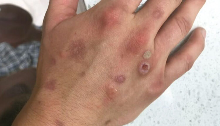
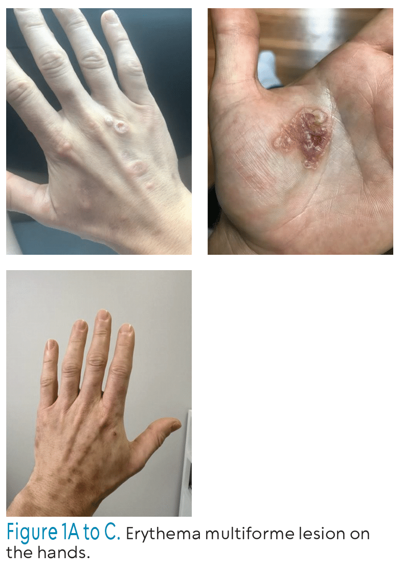
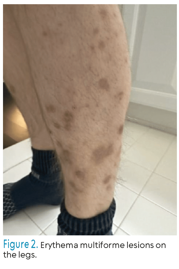
Erythema multiforme (EM) is an acute, immune-mediated dermatologic condition characterized by distinctive target-like lesions of the skin and (Figures 1 and 2), in more severe cases, painful erosions of the mucous membranes. Because EM may involve both cutaneous and mucosal surfaces, nurses play a critical role in comprehensive patient assessment, including evaluation of pain, hydration status, nutritional intake, and the risk for secondary infection. In cases with extensive oral or respiratory involvement, vigilant monitoring for potential airway compromise is essential.
 Effective management of EM often requires interprofessional collaboration (IPC) among nurses, physicians, and oral health professionals.1 Nurses serve as key facilitators of communication between medical and dental teams, particularly when mucosal disease compromises oral health and quality of life.
The World Health Organization defines IPC as a process in which multiple healthcare professionals work together with patients, families, caregivers, and communities to deliver coordinated, patient-centered care.2 This collaborative network commonly includes mental health providers, primary and specialty care physicians, nurse practitioners, registered dietitians, social workers, psychologists,and pharmacists.3,4 Oral health professionals are also essential members of this team, contributing to disease prevention, early detection, and health promotion across the lifespan.
Erythema Multiforme Major and Minor
Erythema multiforme is traditionally categorized into EM minor and EM major, with EM minor defined by typical target lesions with minimal or no mucosal involvement, while EM major includes both target lesions and significant mucosal involvement. These forms of EM are most commonly associated with infectious triggers, particularly herpes simplex virus, and are distinguished by their characteristic raised, targetoid morphology.
 In contrast, mycoplasma pneumoniae–induced rash and mucositis (MIRM) and reactive infectious mucocutaneous eruption (RIME) have emerged as related but distinct post-infectious mucocutaneous syndromes. The classification of MIRM and RIME, and their relationship to EM, remains debated. Some experts consider these conditions to fall within the broader EM spectrum, whereas others regard them as separate entities. Clinically and histopathologically, MIRM and RIME share notable overlap with EM major, particularly in their mucosal involvement, though their cutaneous manifestations are typically sparse or atypical compared with the classic target lesions of EM.
Mycoplasma pneumoniae infection may also precipitate an epidermal necrolysis pattern, and some patients demonstrate overlapping features of EM and Stevens-Johnson syndrome (SJS)/toxic epidermal necrolysis (TEN). Differentiating MIRM/RIME from SJS/TEN is essential because management strategies differ significantly, especially given the more severe course and drug-associated etiology typical of SJS/TEN.
Although EM is most commonly infection related and SJS/TEN is usually drug-induced, the clinical presentation remains the most reliable method of distinguishing these syndromes. EM is defined by typical or atypical raised target lesions, whereas SJS/TEN is characterized by flat purpuric macules that progress to vesicles, bullae, and widespread epidermal detachment. Skin detachment in EM is generally limited, and outcomes are more favorable than in SJS/TEN. Nonetheless, the nosology of these disorders remains complex, as epidermal necrolysis can occur in both EM-related conditions and SJS/TEN, contributing to diagnostic uncertainty.
Rather than relying on the traditional major/minor classification, a more practical approach categorizes acute EM as severe or nonsevere, based on the extent and severity of mucosal involvement. Chronic EM can be severe if it includes prominent mucosal disease (EM major). It is nonsevere if it remains primarily a skin-limited condition (EM minor).
Chronic EM may be recurrent or persistent, often presenting with recalcitrant lesions that intensify during disease flares. Recurrent EM is defined by repeated episodes over several years, with studies reporting an average of approximately six episodes annually and a disease duration of 6 to 10 years. Persistent EM is characterized by the continuous presence of typical or atypical target lesions without periods of complete resolution. Bullous lesions are frequently observed, and mucosal involvement may occur but is not universal.
Diagnostic Confusion with Pemphigus Vulgaris
Pemphigus vulgaris (PV) is a chronic autoimmune mucocutaneous blistering disorder caused by autoantibodies targeting desmogleins, resulting in intraepithelial blister formation. Oral lesions are the initial manifestation in approximately 50% to 70% of cases, and the condition may clinically resemble EM, particularly when painful mucosal erosions are present.
According to the British Association of Dermatologists, management of PV consists of two phases: induction, aimed at controlling disease activity, and maintenance, focused on preventing relapse while minimizing drug exposure.5 Systemic corticosteroid dosing is guided by disease severity, with mild cases treated at 0.5 to 1 mg/kg/day and severe cases requiring up to 2 mg/kg/day.5 Once disease activity is controlled, corticosteroids are tapered gradually.
Systemic steroids are often combined with immunosuppressive agents, such as azathioprine, mycophenolate mofetil, cyclophosphamide, or methotrexate, as well as immunomodulatory therapies including intravenous immunoglobulin or immunoadsorption.6 Oral lesions may be managed with topical corticosteroids or intralesional injections for refractory disease.7
Misdiagnosing PV as EM can significantly affect patient outcomes. EM is typically self-limiting and often requires supportive care, whereas PV necessitates prolonged immunosuppressive therapy. Therefore, thorough clinical evaluation and appropriately performed biopsies remain essential in patients presenting with persistent mucosal lesions.
Medication-Induced Erythema Multiforme
The skin is the organ most frequently affected by adverse drug reactions, occurring in up to 10% of hospitalized patients and approximately 1% to 3% of individuals taking multiple medications. Nearly 30 distinct cutaneous drug reaction patterns have been described, ranging from mild eruptions to life-threatening conditions such as SJS/TEN.
Managing drug-induced skin reactions is particularly complex in older adults with polypharmacy. These cases highlight the need for heightened vigilance, especially when prescribing psychiatric or high-risk medications. Early recognition and prompt discontinuation of the offending agent are essential to prevent progression to severe cutaneous adverse reactions.8
Norimatsu and Norimatsu9 described an elderly woman who developed a drug-induced reaction after initiation of raloxifene for osteoporosis. A drug-induced lymphocyte stimulation test confirmed raloxifene as the causative agent, underscoring the importance of diagnostic testing in complex presentations.
Relationship Between Herpes and Erythema Multiforme
EM is primarily mediated by a cell-mediated immune response, with infections accounting for approximately 90% of cases. Herpes simplex virus (HSV) type 1 is the most common trigger, followed by HSV-2 and mycoplasma pneumoniae, particularly in pediatric populations.
The pathogenic mechanisms differ between infection-triggered and drug-induced EM. In HSV-associated EM, autoreactive T-cells target keratinocytes expressing viral DNA polymerase, resulting in epidermal injury.10,11 In contrast, drug-induced EM is mediated by cytokines such as tumor necrosis factor-α, perforin, and granzyme B, leading to widespread epidermal damage.10,11
Clinically, HSV-associated EM typically presents with classic target lesions, minimal mucosal involvement, and a self-limited course with no reported mortality.10 Drug-induced EM, however, often begins with a flu-like prodrome and progresses to blistering lesions with prominent mucosal involvement.12
External factors, such as trauma and ultraviolet exposure, may also influence lesion distribution, as lesions may appear in previously sunburned areas.13 Early recognition of severe cutaneous adverse reactions is essential to prevent progression to life-threatening conditions such as SJS/TEN.14-16
Photobiomodulation Therapy
Photobiomodulation therapy (PBM) has been utilized in the management of fluconazole‑ induced oral EM, offering a noninvasive approach that supports healing and symptom reduction. Reports describe the laser parameters commonly used, along with positive treatment outcomes such as decreased pain, accelerated lesion resolution, and improved patient comfort. Proposed mechanisms include PBM’s ability to modulate inflammation, enhance cellular repair, and promote tissue regeneration. Practical clinical guidelines emphasize proper laser settings, consistent application protocols, and careful monitoring to ensure safe and effective integration of PBM into EM management.17
Clinical outcomes reported with this approach include immediate and significant pain reduction following the first treatment session, along with marked edema reduction within 48 to 72 hours. Patients typically experience partial mucosal healing within the first few days, ultimately achieving complete resolution of lesions upon completion of therapy.17
Clinical application guidelines for dental practice emphasize initiating PBM therapy as early as possible once EM lesions are identified. A point-by-point technique should be used across all ulcerated and inflamed areas, ensuring consistent energy delivery throughout the treatment sites. Thorough documentation of each session, including wavelength, power, time, spot size, and total energy, is essential for accuracy and reproducibility. PBM should also be combined with supportive measures such as adequate hydration, appropriate analgesics, and careful oral hygiene to optimize healing and patient comfort.17
Analgesic support commonly includes systemic agents such as acetaminophen or non-steroidal anti‑inflammatory drugs to manage discomfort during the acute phase. Oral health professionals should also reinforce oral hygiene instructions, including the use of soft‑bristled toothbrushes, alcohol‑free antimicrobial rinses, and avoidance of spicy or acidic foods, to reduce irritation and promote mucosal healing.
The mechanism of action of PBM involves increasing mitochondrial adenosine triphosphate production, which enhances cellular energy availability. It also reduces pro-inflammatory cytokines and oxidative stress, helping to moderate the inflammatory response.17 Additionally, PBM stimulates fibroblast activity and supports tissue repair while improving microcirculation and promoting angiogenesis, all of which contributes to accelerated healing and improved clinical outcomes.
Interventions and Treatments
Treatment strategies are dictated by the disease’s chronicity (acute vs recurrent) and severity.18 Treatments focus on identifying the cause, managing pain, and preventing secondary infections.
Acute management focuses on symptomatic relief, including topical corticosteroids, antihistamines, and antiseptic or anesthetic washes for painful oral lesions.19,20
Recurrent EM often requires antiviral prophylaxis (eg, acyclovir or valacyclovir) if HSV is the suspected trigger. For those unresponsive to antivirals, second-line therapies include immunosuppressants such as dapsone, azathioprine, or antimalarials.18,19
Recent case studies highlight the success of antimicrobial photodynamic therapy and PBM. These noninvasive laser-based treatments have shown significant improvement in pain reduction and lesion healing within 24 to 48 hours for severe oral EM. 21
Topical steroids are used for acute skin and wound care. Medium-to-high potency topical corticosteroids should be applied to cutaneous (skin) lesions to reduce inflammation and itching. For blistered areas, use nonadherent dressings and saline compresses to soothe the skin and maintain a moist healing environment. Signs of secondary bacterial infection (purulent discharge, foul odor, or increasing heat) should be noted and strict hand hygiene during dressing changes is a must.
For oral and mucosal care, analgesic rinses are helpful. Compounded mixtures containing lidocaine, diphenhydramine, and antacids will numb the area before meals. If oral lesions make swallowing difficult, monitor fluid intake and output and advocate for intravenous fluids if the patient is at risk for dehydration.
If HSV is the confirmed trigger, a nurse will administer antiviral medications (eg, acyclovir). Antihistamines can be used to manage pruritus (an uncomfortable, irritating feeling that encourages scratching of the skin), which can be distressing and leads to skin breakdown from scratching.
In oncology settings, nurses must distinguish EM from toxic erythema of chemotherapy. This requires careful review of the patient’s medication history and the timing of chemotherapy cycles to ensure the correct supportive care is initiated.
Conclusion
EM is a complex mucocutaneous disorder with diverse clinical presentations, fluctuating severity, and a broad range of infectious or medication-related triggers. Understanding the distinctions among EM, MIRM, RIME, and SJS/TEN is essential, as overlapping features frequently lead to diagnostic uncertainty and delays in appropriate treatment. Clear classification based on mucosal involvement and lesion morphology supports timely recognition and helps clinicians differentiate EM from life threatening conditions such as SJS/TEN or autoimmune blistering disorders like PV.
Because EM affects both skin and mucosal tissues, optimal management requires coordinated interprofessional collaboration among nurses, physicians, oral health professionals, dermatology specialists, and allied health professionals. Nurses play a particularly critical role through early assessment, airway monitoring, wound care, medication management, patient education, and identification of red flag symptoms that suggest severe disease progression. Complex cases, including chronic, recurrent, or persistent EM, often require long-term antiviral therapy, immunomodulatory regimens, and ongoing reassessment to prevent flares and improve quality of life.
Emerging therapies, such as PBM and antimicrobial photodynamic therapy, offer promising adjunctive options for patients with painful oral lesions, reflecting broader advancements in supportive dermatologic care. Additionally, attention to medication safety, especially in older adults with polypharmacy, remains essential to reducing the risk of drug induced EM and more severe cutaneous adverse reactions.
Ultimately, improving outcomes for patients with EM depends on three pillars: accurate and timely diagnosis, evidence-based therapeutic strategies tailored to disease severity and chronicity, and effective interprofessional communication across all points of care. Continued research is needed to further define the immunologic mechanisms of EM, refine diagnostic criteria, and develop targeted therapies that address both the cutaneous and mucosal manifestations of this multifaceted condition.
References
- Hung M, Birmingham WC, Tucker M, Schwartz C, Mohajeri A. Integrating dentistry into interprofessional healthcare: a scoping review on advancing collaborative practice and patient outcomes. Healthcare. 2025;13(21):2780.
- World Health Organization. Framework for Action on Interprofessional Education Collaborative Practice. Available at who.int/publications/i/item/framework-for-action-on-interprofessional-education-collaborative-practice. Accessed August 21, 2026.
- Rawlinson C, Carron T, Cohidon C, et al. An overview of reviews on interprofessional collaboration in primary care: barriers and facilitators. Int J Integr Care. 2021;21:32.
- Kattach L, Singleton H, Ersser S, Holley D, Pearson I, Shadeed A. Nurse-led models of service delivery for skin cancer detection: a systematic review. J Adv Nurs. 2025;81:8432-8456.
- Harman KE, Brown D, Exton LS, et al. British Association of Dermatologists’ guidelines for the management of pemphigus vulgaris. Br J Dermatol. 2017;177:1170–1201.
- Kridin K, Zelber-Sagi S, Bergman R. Pemphigus vulgaris and pemphigus foliaceus: differences in epidemiology and mortality. Acta Derm Venereol. 2017;97:1095–1099.
- Al-Harbawee A, Kassam K, Patel AN, Cottom H, Cheng L. Oral pemphigus vulgaris: dentists take-home message. Clin Case Rep. 2021;9:e04494.
- Rezapour, M., Mesgarankarimi, A. Low-dose venlafaxine-induced erythema multiforme: a case report. J Med Case Reports. 2025;19:188 (2025).
- Norimatsu Y, Norimatsu Y. First report of erythema multiforme minor caused by raloxifene hydrochloride. Case Rep Dermatol. 2021;13:445–449.
- Aurelian L, Ono F, Burnett J. Herpes simplex virus (HSV)-associated erythema multiforme (HAEM): a viral disease with an autoimmune component. Dermatol Online J. 2003;9:1.
- Ono F, Sharma BK, Smith CC, Burnett JW, Aurelian L. CD34+ cells in the peripheral blood transport herpes simplex virus DNA fragments to the skin of patients with erythema multiforme (HAEM). J Invest Dermatol. 2005;124:1215–1224.
- Samim F, Auluck A, Zed C, Williams PM. Erythema multiforme: a review of epidemiology, pathogenesis, clinical features, and treatment. Dent Clin North Am. 2013;57:583–596.
- Huff C, Weston WL. The photodistribution of erythema multiforme. Arch Dermatol. 1980;116:477.
- Demoly P, Adkinson NF, Brockow K, et al. International Consensus on drug allergy. Allergy. 2014;69:420–437.
- Sassolas B, Haddad C, Mockenhaupt M, et al. ALDEN, an algorithm for assessment of drug causality in Stevens-Johnson Syndrome and toxic epidermal necrolysis: comparison with case-control analysis. Clin Pharmacol Ther. 2010;88:60–68.
- Lazarević VV. Diagnosis, investigation and management of non-immediate (type iv) cutaneous adverse drug reactions. Acta Dermatovenerol Croat. 2024;32:88–95.
- Silva de Souza F, Palma VM, Lonni N, Rabelo GD. Photobiomodulation therapy as an effective approach for fluconazole-induced oral erythema multiforme: a case report. Lasers Dent Sci. 2026;10:4.
- Soares A, Sokumbi O. Recent updates in the treatment of erythema multiforme. Medicina. 2021; 57:921.
- Hafsi W. Erythema multiforme. Available at ncbi.nlm.nih.gov/books/NBK470259. Accessed August 21, 2026.
- Copp LB. Erythema multiforme. Journal of the Dermatology Nurses’ Association. 2024;16(6):224–227.
- Martins F, Pallos D, Candeia J, Zerbinati R, Braz-Silva PH, Campos L. Noninvasive techniques for management of erythema multiforme. Case Rep Dent. 2023;2023:9938939.
From Dimensions of Dental Hygiene. September/October 2026; 24(5):40,43-45