
Furcation Anatomy
Correctly assessing furcations begins with an understanding of their basic structure.
 Assessing furcations is one of the many clinical parameters of the periodontal evaluation. It is a topic with many facets, but this feature will focus on the root morphology of furcation anatomy. In health, this area is covered by gingiva and alveolar bone. With progressive loss of attachment, furcation involvement occurs. Knowledge of the location of furcations allows the clinician to assess and diagnose a patient’s periodontal condition, choose the appropriate instruments for debridement, provide a patient with the best oral hygiene aid to access the site for daily biofilm removal, and refer a patient to a periodontist as indicated.1-3
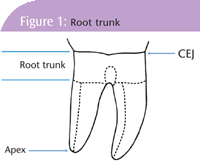
Teeth typically have one, two or three roots. The anatomic root extends from the cemento enamel junction (CEJ) to the apex and is covered by cementum. The root trunk extends from the CEJ to the entrance of the furca (see Figure 1). Anterior teeth generally have one root. The maxillary first premolar has a buccal and lingual root (61% of the time)4 with two furcation entrances (mesial and distal). Additionally, this tooth has a mesial concavity that extends from the crown onto the middle third of the root trunk.4 Mandibular molars have two roots (mesial and distal) with two furcation entrances (buccal and lingual). Maxillary molars have three roots (mesiobuccal, distobuccal and palatal) with three furcation entrances (buccal, mesial and distal).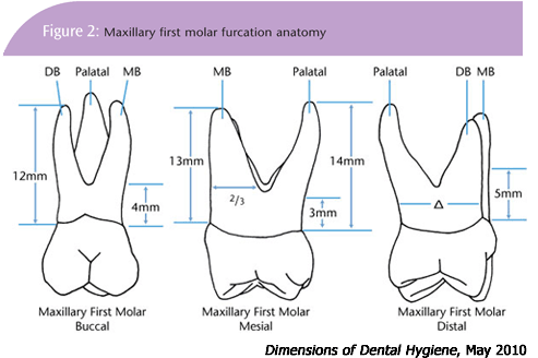
PLANE CONSIDERATIONS
Several studies4-9 have been conducted to determine the location of furcations. When assessing location, the clinician must take into account both the horizontal and vertical planes.
On the buccal aspects of maxillary first molars, the average distance from the CEJ to the furcation is 4 mm and is centered mid-facial. The mesial furcation is 3 mm from the CEJ and is not centered. Because the mesio-buccal (MB) root is so large, the mesial furcation is usually found about two-thirds of the way in a buccal (B) lingual (L) direction toward the palate. Thus, it should be accessed from the lingual. The distal (D) furcation is located 5 mm from the CEJ and is centered. Therefore, it can be accessed from either the buccal or the lingual—although the lingual embrasure is larger, so the lingual approach may still be easier. (Figure 2.)
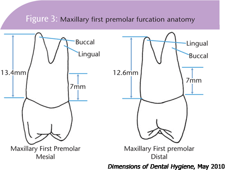
The maxillary first premolars (that are bifurcated) have furcations that are midmesial and mid-distal, and are both located 7 mm from the CEJ. (Figure 3.)
The furcations on the mandibular first molars are centered mid-facial and midlingual. The average distance from the CEJ to the furca is 3 mm on the buccal and 4 mm on the lingual. (Figure 4.) The positions of furcations differ on second and third molars.

The root trunks on second and third molars are longer and the roots are more fused.
Furcation involvement may be measured and classified using a Nabors probe. Furcation involvement may be detected clinically before there is evidence radiographically.
ASSESSING FURCATIONS
The closer the furcation is to the CEJ, the easier it is for both the clinician and patient to access. These are generally found on first molars. The further the furcation is from the CEJ, the less likely it is to become periodontally involved. A minimum of 5 mm of attachment loss on molars may result in early furcation involvement.10 Horizontal bone loss and attachment loss of 6 mm or more would result in Class II furcations on both maxillary and mandibular first molars.5-6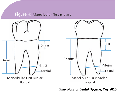
Second and third molars have longer root trunks and their furcations are located more apically. The more apical the furcation the more difficult it is to access and treat.
Knowing the location of furcations allows the clinician to: assess and document furcations; determine which instruments are the best choice for debridement; provide home care instructions; and recommend referral as appropriate.
REFERENCES
- Cooper MD, Mann NK. Instrumentation for root debridement. Contemporary Oral Hygiene. 2007;7(2):30-35.
- Nield-Gehrig J. Advanced instrumentation techniques for root surface debridement. Journal of Practical Hygiene. 2004;13(3):19-22.
- http://www.perio.org/resources-products/pdf/
management.pdf. Accessed August 25, 2009. - Scheid, RC. Woelfel’s Dental Anatomy Its Relevance to Dentistry. 7th ed. Philadelphia, PA: Lippincott Williams & Wilkins, 2007:205.
- Gher MW, Dunlap RW. Linear variation of the root surface area of the maxillary first molar. Journal of Periodontology. 1985;56(1):39-43.
- Dunlap RW, Gher MW. Root surface measurements of the mandibular first molar. Journal of Periodontology. 1985;56(4):234-238.
- Plagmann HC, Holtorf S, Kocher T. A study of the imaging of complex furcation forms in upper and lower molars. Journal of Clinical Periodontology. 2000;27:926-931.
- Santana RB, Uzel MI, Gusman H, et al. Morphometric Analysis of the Furcation Anatomy of Mandibular Molars. Journal of Periodontology. 2004;75(6):824-828.
- McKechnie LB. Root morphology in periodontal therapy. Dental Hygienist News. 1993;6(1);3-6,22-3.
- Nield-Gehrig, J. Fundamentals of Periodontal Instrumentation. 6th ed. Philadelphia, PA: Lippincott Williams & Wilkins. 2008:458.
From Dimensions of Dental Hygiene. May 2010; 8(5): 36, 38-39.

I lovvved the illustrations and the diagrams. I wonder if I can use them for my students?